Miniature Imaging Tools Enable Real-Time Neural Observation During Natural Behavior
According to Bioengineer.org, researchers have developed a miniature microscope that can observe and control brain cells while an animal moves naturally.
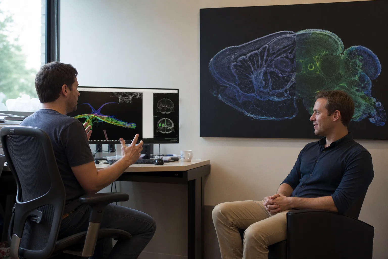
The report is potentially important for neural-circuit research because it points toward a less static view of brain activity—one that follows cells during behavior rather than isolating the brain from movement. At this stage, however, the available report provides no technical specifications, animal model, cell type, or details of the control method.
A smaller instrument, a more natural question
Many neuroscience experiments become visually and biologically cleaner when movement is restricted. That control can help researchers separate signals, but it also changes the conditions under which neural circuits operate. The headline reported by Bioengineer.org describes a different experimental aim: observing and manipulating brain cells during natural movement.
The distinction matters. A neural signal recorded in a stationary preparation is not automatically equivalent to one produced while an animal is navigating its environment. Movement changes the sensory stream, the timing of motor commands, and the coordination between brain regions. A tool that can preserve this behavioral context could therefore make it easier to examine how neural activity unfolds as part of an intact action.
The central visual challenge is equally important. A microscope must capture cellular activity with enough clarity to distinguish meaningful changes in the visual field, while remaining small enough not to prevent the movement researchers want to study. In practical terms, the instrument is being presented as both an imaging system and a way to influence selected brain cells.
That combination is more consequential than observation alone. Recording can reveal correlations between cells and behavior; control can test whether a particular activity pattern is functionally involved. The evidence available here does not establish how precise that control is, or whether the reported system has already demonstrated a specific behavioral effect. Those details will determine the method’s real value.
What the report does—and does not—establish
The confirmed information supports a narrow conclusion: a miniature microscope is associated with the ability to observe and control brain cells during natural movement. It does not identify the researchers, the laboratory, the species used, the scale of the device, or the neural activity being measured. It also does not state whether the work concerns zebrafish, mammals, or another model organism.
For readers following neural network formation and zebrafish neurobiology, that missing context is not a minor editorial gap. The usefulness of a miniature imaging system depends on the animal’s size, transparency, movement pattern, and the accessibility of its nervous system. It also depends on the spatial and temporal resolution of the recordings—details that are not included in the available source material.
The safest interpretation, then, is not that neuroscience has gained a universal window into the moving brain. It is that a reported instrument may help shift experiments toward behaviorally natural conditions. The next information to check is the original study: which cells were targeted, how movement was preserved, what “control” meant experimentally, and whether the method was validated against an established imaging approach.
For visual neuroscience, the practical principle is simple: do not judge a new microscope by its miniature scale alone. Judge the relationship between the image it produces, the movement it permits, and the causal question it can actually answer.