Zebrafish brain imaging: 5 ways to prevent motion artifacts
During a fast volumetric calcium scan, a larval zebrafish twitches — a reflex adjustment, a momentary flex in a quiet dish of E3 medium. The displacement is small, submillimeter, almost invisible to the naked eye.
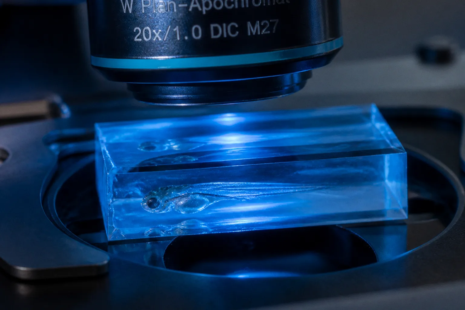
To the microscope, that twitch becomes a streak of false signal smeared across an entire functional brain map. Every calcium transient, every neural population burst the researcher meant to attribute to a stimulus, now carries the ghost of locomotion baked into the data.
Motion artifacts are not a minor inconvenience in zebrafish brain imaging. They are the central bottleneck between a beautifully resolved brain and a quantifiable one. The good news is that the bottleneck is engineered, not intrinsic. Five distinct strategies — chemical, mechanical, microfluidic, genetic, and computational — each attack a different layer of the problem. The art of a clean imaging session is choosing the right combination without suppressing the biology the experiment was designed to measure.
Beyond Tricaine: Chemical Paralysis and Neuromuscular Blockers
For two decades, MS-222, or tricaine, was the default anesthetic on the zebrafish rig. It works quickly, it is inexpensive, and many introductory protocols begin with the same 0.016% working solution. The problem is not that tricaine fails to immobilize a larva. It does. The problem is how it does so: tricaine blocks voltage-gated sodium channels throughout the nervous system, which means it suppresses the very action potentials a functional brain imaging experiment is trying to record. Heart rate drops. Hemodynamic responses shift. The brain being imaged is no longer quite the brain intended for study.
For functional calcium imaging, the practical substitute is a non-depolarizing neuromuscular blocker. Pancuronium bromide, applied at 300 µM (0.3 mg/ml) in the imaging medium, paralyzes skeletal muscle without crossing into the central nervous system at meaningful concentrations. The larva stops moving while the brain continues to generate activity. GCaMP6f transients remain more interpretable than they would under a centrally acting anesthetic. This is the first lever many labs pull when motion artifacts contaminate a session, and it remains a common pharmacological intervention in larval functional imaging.
The trade-off is real, though often manageable. Pancuronium is not free of physiological cost. Prolonged exposure can alter heart rate and respiratory patterning, and paralysis removes any possibility of using tail movements as a behavioral readout during the same recording. That last point is easy to overlook when a protocol combines several otherwise sensible steps: a tail may be physically exposed by an agarose mount, but it cannot provide a motor signal if pancuronium has blocked neuromuscular transmission.
For short imaging windows — typically under two hours — the effect may be tolerable, depending on the preparation and the endpoint. For longer experiments, especially those tracking development across days, the researcher must weigh artifact reduction against the introduction of a new confound. Some groups alternate paralysis sessions with brief recovery windows. Others run separate behavioral and imaging cohorts: one preparation is mounted to preserve tail or eye movements, while another is chemically paralyzed for a stable neural recording. Still others move directly to genetic immobilization, which sidesteps repeated pharmacological exposure altogether.
Comparing paralysis agents for functional imaging
| Parameter | Tricaine (MS-222) | Pancuronium bromide |
|---|---|---|
| Mechanism | Blocks voltage-gated Na⁺ channels centrally | Non-depolarizing nicotinic receptor antagonist at the neuromuscular junction |
| Effect on brain activity | Strongly suppresses action potentials | Minimal direct central effect at working concentrations |
| Typical working concentration | 0.016% | 300 µM (0.3 mg/ml) |
| Suitability for functional imaging | Poor when native neural dynamics are the endpoint | Good for recordings that require skeletal-muscle paralysis |
| Primary use case | Anesthesia during dissection, mounting, and terminal procedures | Paralysis during functional calcium imaging |
| Behavioral readouts during exposure | Suppressed by anesthesia | Not available from paralyzed skeletal muscle |
The practical distinction is not simply “which drug immobilizes the fish?” It is “which part of the animal must remain functional?” If the experiment measures brain activity alone, pancuronium may be an efficient first-line intervention. If it measures the relationship between neural activity and locomotion, the preparation must preserve motor output — or the behavioral and imaging measurements must be separated in design.
Optimizing Physical Embedding with LMP Agarose and Stamping Devices
Before any chemical enters the dish, most imaging sessions begin with a physical decision: how is the larva held in place? Low-melting-point agarose, typically at 1.2% to 1.8% in E3 medium, is the workhorse. The larva is drawn into a drop of cooled agarose on a glass-bottom dish, oriented under a stereoscope, and left to set. Within minutes, the body is encased in a clear gel that holds the head stationary for the duration of the scan.
The advantage is optical clarity. LMP agarose matches the refractive index of water closely enough that two-photon and light-sheet excitation can penetrate without severe aberration. It is also cheap, forgiving, and easy to adapt when the field of view changes. A researcher can mount the brain dorsally for whole-brain activity, rotate the animal for a lateral view, or adjust the head angle to fit an objective with a narrow working distance.
The disadvantage is mechanical: agarose restricts the entire animal unless the researcher deliberately cuts it away. That means the eyes cannot move, the tail cannot flick, and any stimulus that depends on motor feedback — a virtual-reality corridor, a looming dot assay, or an optomotor response paradigm — becomes impossible in a fully embedded preparation. The solution is not to pretend that every readout survives immobilization. It is to design the mount around the readout.
After the agarose sets, fine forceps or a small tungsten needle can remove the gel around selected structures. In a preparation intended for eye tracking, the eyes may be exposed while the head remains stable. In a behavior-compatible mount, the tail can be released so that it is available for movement recording. That arrangement belongs to a behavioral imaging workflow, however. If pancuronium is added afterward, the exposed tail may be visible but it will not provide a meaningful motor output. Physical access and physiological function are separate variables.
A more recent approach replaces the manual embedding ritual entirely. The Agarose Stamped Device (ASD) is a low-cost platform in which larva-sized wells are molded into a thin agarose sheet using a 3D-printed stamp. Larvae are drawn into the wells by surface tension, oriented in parallel, and held in place without individual handling. Depending on the design and the experiment, eye, mouth, and tail can remain accessible. For screens involving dozens of larvae — pharmacological, developmental, or high-content behavioral — the ASD turns an hour of manual mounting into a few minutes of stamping, while reducing the orientation variability that plagues hand-embedded samples.
The device does not eliminate the need for judgment. A stamped well can standardize position, but it cannot decide whether the experiment needs the fish fully restrained, partially exposed, or free to generate a motor response. Nor does a uniform mount solve every optical problem: the brain still has to sit at the correct depth, the agarose must remain clear, and pressure points must not distort the larva. The value of the ASD is that it makes those decisions repeatable.
This is the kind of method that rewards patience with throughput. A lab running ten larvae per condition may manage with hand mounting. A lab running a hundred larvae per condition can no longer afford to treat orientation as an artisanal step.
Microfluidic Restriction: The ZEBRA Platform for Automated Positioning
Agarose is not the only way to hold a larva still. The Zebrafish Entrapment By Restriction Array, or ZEBRA, takes the immobilization problem out of the gel and into a microfabricated channel. Larvae are loaded into a PDMS device where narrow constrictions match the diameter of the animal at several points along its body. The fish cannot turn or flex, but remains bathed in circulating medium. Waste diffuses away, fresh buffer flows in, and temperature and oxygenation can be maintained for extended recordings.
The appeal is reproducibility. Every larva in a ZEBRA chip sits at a comparable angle, depth, and distance from the objective. Image registration becomes easier before any computational correction begins. Stimulus delivery, whether pharmacological or sensory, can be timed through channel switching. For experiments that demand standardization — screens of mutant libraries, dose-response curves, or longitudinal imaging across many animals — ZEBRA removes a substantial fraction of the variance introduced by hand embedding.
Microfluidics also changes the logic of the experiment. In agarose, the animal is immobilized by a solid material surrounding its body. In a restriction device, the animal is held by geometry while the surrounding medium remains exchangeable. That can make perfusion-based protocols more straightforward and can reduce the delay between changing the external environment and exposing the larva to a new condition. It also makes the flow itself part of the system: pressure, shear, bubble formation, and channel blockage become variables that need monitoring.
The trade-off is fabrication. ZEBRA chips require cleanroom access or a soft-lithography setup, which places the technique beyond the reach of labs without microfabrication infrastructure. Once fabricated, the chips are reusable and the per-experiment cost is low, but the entry cost is real. Loading can also be less forgiving than placing a single larva in agarose. A fish that enters at the wrong angle may be difficult to reposition without restarting the preparation.
The practical question for a group considering the platform is throughput: at what point does manual agarose mounting become the limiting step in the pipeline, and is that point close enough to justify the fabrication overhead? For a one-off, highly customized preparation, agarose remains difficult to beat. For repeated experiments where position and perfusion must be comparable from animal to animal, a microfluidic restriction array starts to look less like specialized hardware and more like basic infrastructure.
A larva that cannot move is not yet a controlled experiment. The constraint is only useful if it preserves the brain you wanted to record.
Genetic Immobilization via Alpha-Bungarotoxin Expression
Pharmacological paralysis is reversible, repeatable, and well understood. It is also external: applied from a pipette, washed out at the end of the session, and absent when the researcher wants the animal to recover. For experiments that require immobilization from fertilization through several days of development, the dose-response cycle becomes impractical.
Genetic immobilization solves this by building the paralysis into the animal. Alpha-bungarotoxin, a peptide derived from snake venom that irreversibly blocks nicotinic acetylcholine receptors at the neuromuscular junction, can be expressed endogenously via mRNA injection at the one-cell stage. The toxin is produced by the embryo itself, acetylcholine transmission is blocked from the earliest stages of motor development, and the larva remains paralyzed through the developmental window of interest.
Concentrations around 2 µM in the relevant tissues have been used to achieve strong immobilization with minimal reported developmental side effects. The method is especially valuable for long-term functional imaging sessions where daily pharmacological dosing would be impractical. It also removes one source of session-to-session variation: the researcher is not repeatedly deciding whether a larva has received enough blocker, whether wash-in is complete, or whether a changing bath concentration is contributing to changes in physiology.
The advantages are striking. There is no daily dosing, no anesthetic wash-in, and no cumulative drug exposure from repeated treatment. Imaging sessions can run across several developmental stages without interrupting the preparation for drug administration. For experiments that compare brain activity across developmental stages within the same animal, genetic immobilization can provide a consistent baseline that chemical paralysis is less well suited to provide.
But the central limitation is equally clear: the animals cannot move at any point. That forecloses experiments with a motor readout, not merely during the imaging interval but throughout the period in which the construct is active. Alpha-bungarotoxin expression must also be verified for each line or injection strategy. Mosaic expression can produce partially mobile larvae that defeat the purpose of the method and introduce an awkward intermediate phenotype: enough movement to create artifacts, but not enough to count as a useful behavioral preparation.
Potential effects on sensory neurons that also express nicotinic receptors deserve attention as well. Even when the primary target is the neuromuscular junction, a genetic manipulation that alters cholinergic signaling may change more than motor output. The magnitude of such effects depends on the expression strategy and experimental context, so the appropriate control is not simply an untreated larva. Researchers should compare neural activity and developmental morphology in immobilized and control animals using the same imaging conditions.
The decision rule is straightforward. If the experiment ends when the larva moves, pharmacological paralysis is faster and cheaper. If the experiment requires the larva to remain still throughout a developmental window while recording continues, genetic immobilization is the more faithful tool. If the experiment needs both long-term neural imaging and behavior, the answer may be two complementary preparations rather than one compromise animal.
Computational Restoration: TMAC and NoRMCorre Algorithms
Even with optimal physical and chemical stabilization, residual motion persists. A beating heart can produce micrometer-scale displacements of the brain every few hundred milliseconds. Slow changes in agarose tension shift the field of view over minutes. Stage noise from the microscope itself adds subpixel jitter to every frame. None of these movements is necessarily large enough to ruin an image, but each can corrupt a calcium trace.
NoRMCorre addresses this with piecewise rigid motion correction. The imaging field is divided into overlapping spatial patches; each patch is registered to a reference frame, and the resulting displacement field is smoothed across the whole image. Subpixel lateral and axial motion can be corrected frame by frame, and the algorithm runs quickly enough to keep pace with volumetric calcium imaging at high frame rates.
The important word is “piecewise.” NoRMCorre is well suited to translation and local rigid displacement, but it is not a magic eraser for tissue deformation. If the brain changes shape as the heart beats, or if different structures move independently, a rigid model can leave behind residual distortion. That does not make the method inadequate. It means the researcher should inspect the corrected data rather than trusting a registration score alone.
TMAC takes a different approach. It assumes the researcher is recording two channels simultaneously: one activity-dependent, such as GCaMP, and one activity-independent, such as RFP or a similar fluorophore expressed under the same promoter. The activity-independent channel provides a reference for motion and noise rather than neural signal. A generative model learns the relationship between the channels and estimates the motion component in the activity-dependent signal, allowing that component to be removed from the calcium trace.
The advantage over rigid registration is that TMAC can address non-rigid distortions: the slight warping of tissue as the heart beats, or the small deformations that piecewise rigid correction cannot fully model. The cost is experimental design. A second channel must be available, it must be sufficiently bright and stable, and its expression must be biologically independent of the activity fluctuations being measured. A weak reference channel is not a neutral inconvenience; it can make the correction unstable or encourage the model to remove genuine signal.
For the researcher deciding between the two, the question is therefore not which algorithm is universally better. It is what information the dataset contains. If the pipeline already expresses a second fluorophore in every animal, TMAC offers a route to correcting non-rigid motion without changing the mounting protocol. If the experiment is single-channel, NoRMCorre is the reliable default for residual drift, stage noise, and modest local displacement. Neither replaces the need for good immobilization. Both make good immobilization better.
Computational correction should also happen early enough to inform experimental decisions. A few representative raw stacks can reveal whether the main problem is sudden displacement, slow drift, axial movement, or tissue deformation. Those failure modes point to different interventions. Strong frame-to-frame jumps suggest a physical restraint problem. A gradual shift may indicate mounting tension or stage drift. Periodic deformation synchronized with the heartbeat may be better addressed through a reference channel or a more appropriate registration model.
Practical comparison of the five motion-control strategies
| Method | Best for | Typical setup | Key limitation |
|---|---|---|---|
| Pancuronium bromide paralysis | Short functional imaging sessions | 300 µM in imaging medium | Cardiac effects may become more important over longer exposures; motor readouts are unavailable |
| LMP agarose and stamped devices | Flexible mounting and higher-throughput imaging | 1.2–1.8% agarose or stamped wells | Full embedding prevents behavior unless selected structures are deliberately exposed |
| ZEBRA microfluidic mount | Standardized, automated positioning and perfusion | PDMS chip with channel constrictions | Requires fabrication infrastructure and careful loading |
| Genetic α-bungarotoxin expression | Long-term developmental imaging | mRNA injection at the one-cell stage | Irreversible; motor readouts are not possible |
| TMAC or NoRMCorre | Residual motion after physical stabilization | Computational processing of raw image stacks | Cannot compensate for every form of movement or replace a stable preparation |
The five methods are not alternatives. They are layers. A clean functional dataset is what you get when every layer is doing its job.
Choosing the Stack
The five techniques are not redundant. They operate at different stages of the imaging pipeline, and a well-designed experiment usually combines at least three of them. The correct combination depends first on what must remain measurable.
A session devoted exclusively to neural activity might use pancuronium bromide together with a fully stabilizing LMP agarose mount, followed by NoRMCorre correction on the resulting stack. This is a coherent motion-control strategy because the tail is not being used as a behavioral output. The chemical blocker handles skeletal-muscle movement, the agarose limits gross displacement, and the computational step removes residual drift and local jitter.
A behavior-linked experiment requires a different arrangement. The larva may be mounted in agarose with the tail or eyes deliberately exposed, but pancuronium should not be added if tail movement is the readout. In that case, the researcher must accept some residual motion, use a mounting geometry that stabilizes the brain without eliminating the relevant behavior, or separate the neural imaging and behavioral measurements into different preparations. A physically exposed tail is not equivalent to a functional tail under neuromuscular blockade.
For a lab setting up its first functional imaging rig, the simplest viable stack for brain-only recording is pancuronium bromide in LMP agarose, processed through NoRMCorre. Every component is relatively accessible, the workflow is easy to modify, and the failure modes are visible. The first control should be a recording that includes the reference channel or structural signal needed to distinguish real neural transients from movement. Without that comparison, a clean-looking trace can still be a motion artifact.
For a lab scaling to dozens of larvae, the ASD or ZEBRA platform replaces manual mounting with parallel positioning. The choice between them follows the workflow: stamped agarose offers a lower-complexity route to standardized placement, while microfluidics provides tighter control over position and medium exchange. Neither should be selected only because it increases throughput. The gain matters when the experiment is already bottlenecked by mounting, not when the real limitation is imaging time, data storage, or analysis.
For a lab running longitudinal developmental studies, genetic immobilization removes the daily pharmacological cycle and can make repeated imaging more consistent. It also changes the biological question by eliminating motor output across the developmental window. That trade-off must be written into the experimental design rather than treated as a technical footnote.
For a lab pushing the limits of signal-to-noise, TMAC paired with a second fluorophore can turn residual non-rigid motion into a tractable modeling problem. NoRMCorre remains valuable for the simpler components of drift and translation. In practice, the strongest pipeline may use both: registration to align frames, followed by a channel-informed correction for distortions that alignment alone cannot resolve.
The deeper principle is that immobilization is not a single decision. It is a stack. The researcher chooses how to constrain the animal at the chemical level, how to constrain it at the mechanical level, and how to clean up the residue computationally. Each choice trades off against the others: pancuronium buys stillness but removes motor readouts; agarose buys stability but can block behavior; α-bungarotoxin buys long-term consistency but eliminates movement entirely; microfluidics buys reproducibility at the cost of fabrication; computational correction buys recovery from residual artifacts but cannot recover information that was never recorded cleanly.
The art of the experiment is choosing the stack that preserves the variable actually being measured. A zebrafish brain imaging setup should not be judged by how completely it stops the animal. It should be judged by whether it stops the unwanted movement while leaving the relevant physiology intact.
All five approaches converge on the same goal: a brain that can be recorded without the recording itself disturbing what it measures. Motion artifacts are not a research inconvenience to be tolerated. They are a controllable variable. Treat them as such, and the zebrafish brain gives up its activity with the clarity the experiment deserves.