Zebrafish immobilization: choosing the right imaging method
Your calcium movie looks clean until you ask the uncomfortable question: are those neural signals from the larva, or from the immobilization method?
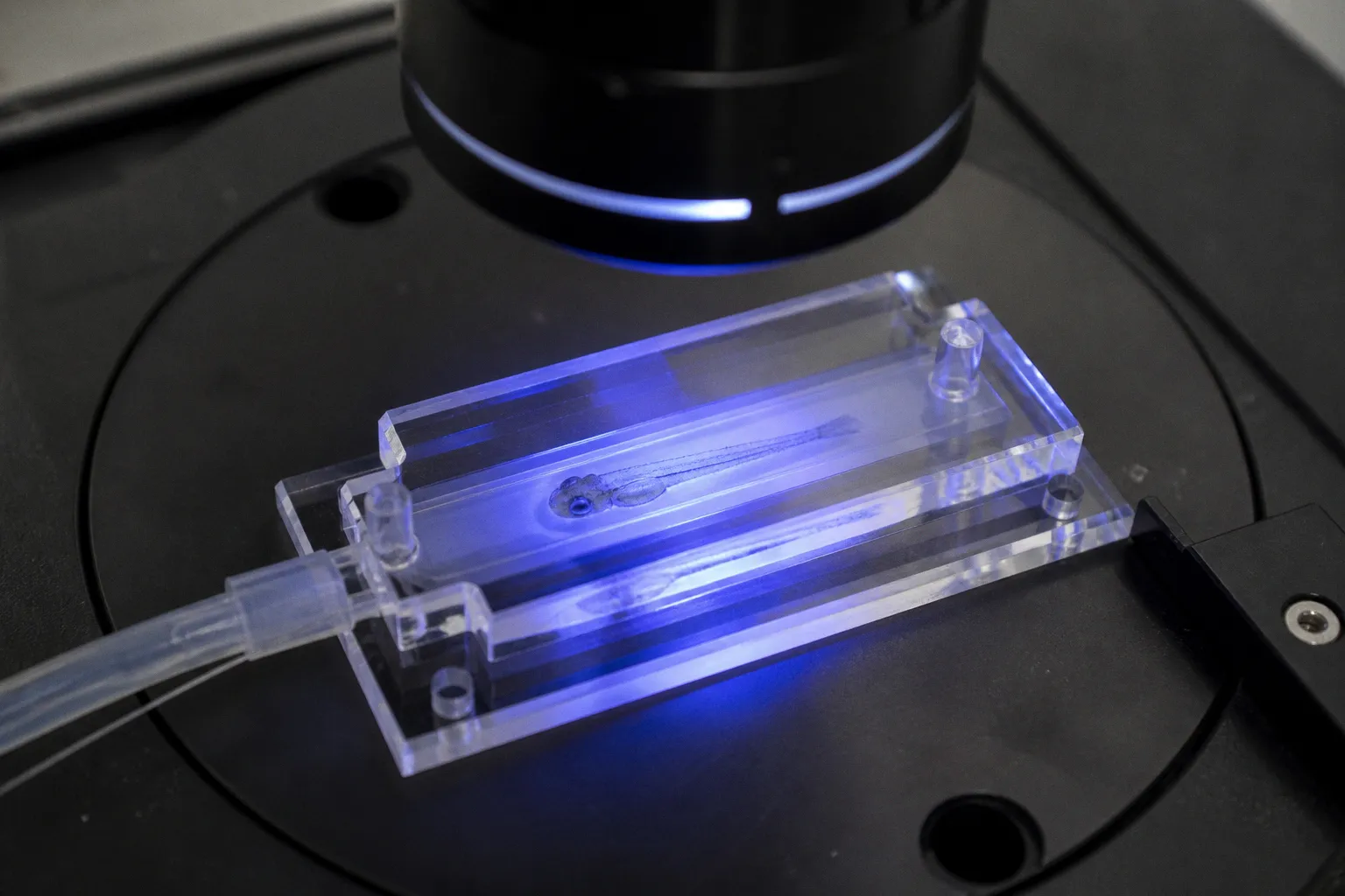
This is where many zebrafish imaging experiments quietly go off the rails. The fish stops moving, the brain looks beautifully still, and the microscope produces a convincing stream of fluorescent activity. But if you used Tricaine to achieve that stillness, you may also have suppressed the very voltage-dependent activity and intracellular calcium oscillations you wanted to measure. A motionless larva is not automatically a physiologically useful larva. Sometimes it is simply a larva whose nervous system has been chemically turned down.
Compare car rental deals in United Kingdom
See available offersPartner link — DiscoverCars comparisonChoosing among zebrafish immobilization methods for calcium imaging means balancing three competing demands: physical stability, preserved neural activity, and acceptable physiological health over the length of the experiment. Let’s walk through the main options and where each one starts to misbehave.
The Tricaine trap: convenient, familiar, and wrong for many functional recordings
Tricaine, or MS-222, remains a familiar tool in zebrafish laboratories because it works quickly, is easy to prepare, and produces obvious paralysis. For routine handling, morphological imaging, or procedures where neural activity is not the readout, that convenience can be perfectly reasonable.
Functional calcium imaging is a different animal.
Tricaine blocks voltage-gated sodium channels. Those channels support action-potential generation and propagation, so blocking them can suppress neural activity and reduce intracellular calcium oscillations. In other words, Tricaine does not merely stop the tail from flicking while leaving the brain untouched. It changes the electrical conditions under which the circuit operates.
That distinction matters most when you are imaging spontaneous activity, sensory responses, escape-related circuits, motor networks, or any behavior-linked pattern. If the neural event depends on action potentials, Tricaine can erase, weaken, or reshape the signal before your objective ever sees it.
The problem becomes even less attractive during early development. Exposure to 30 µg/ml Tricaine between 20 and 48 hours post-fertilization has been associated with developmental delays of roughly 2–6 hours. At concentrations as low as 100 µg/ml, Tricaine can induce pericardial edema. Those effects do not make every use of MS-222 unacceptable, but they do make it a poor default for experiments that depend on intact physiology.
If your experiment measures neural activity, do not treat “the fish stopped moving” as proof that your immobilization worked. It may only prove that the anesthetic reached the brain.
What if you already collected a Tricaine dataset?
First, do not throw the entire experiment into the chemical-waste bin. Ask what the experiment was actually measuring.
If you captured gross anatomy, reporter expression, cell counts, or a structural endpoint, Tricaine may not invalidate the dataset in the same way it would invalidate a functional recording. If you measured calcium transients, however, the interpretation needs much more caution. Compare signal amplitude, event frequency, baseline fluorescence, and stimulus-evoked responses against untreated or alternative-immobilization controls.
A particularly useful control is a preparation in which the larva receives physical immobilization or a neuromuscular blocker instead of Tricaine, while the imaging settings and stimulus remain unchanged. The goal is not to prove that one condition is “normal” in an absolute sense. The goal is to find out whether Tricaine changes the neural readout relative to a method that does not block central voltage-gated sodium channels.
For in vivo calcium imaging, that comparison is not a luxury. It is part of method validation.
Neuromuscular blockers: stop the body without silencing the circuit
Neuromuscular blockers offer a more targeted solution. Rather than blocking voltage-gated sodium channels throughout excitable tissue, they interfere with transmission at the neuromuscular junction. The larva cannot execute a muscle contraction, but the brain remains available for recording.
Pancuronium bromide is one commonly used option, typically at 300 µM in larval preparations. It acts as a non-depolarizing neuromuscular blocker by targeting nicotinic acetylcholine receptors at the neuromuscular junction. That gives you the basic experimental separation we want:
- the muscles cannot generate the movement that would blur the image;
- the central nervous system can continue producing electrical activity;
- calcium signals remain available for functional imaging.
This is not magic paralysis, and the microscope will not reward sloppy prep simply because you changed compounds. Pancuronium can still introduce physiological effects that deserve controls, particularly during long imaging sessions. The long-term impact of chronic exposure on larval brain development remains incompletely documented, so avoid treating it as a universally neutral substitute for Tricaine.
Still, for many active-circuit experiments, it is a more logical starting point.
The practical decision: what must remain alive in your assay?
Before selecting a blocker, write down the part of the animal that must remain functionally responsive.
If you need spontaneous brain-wide activity, the central neural circuit must remain active. If you need a visual response, the eyes and downstream visual pathways must receive and process the stimulus. If you need a tail movement as the behavioral output, complete paralysis defeats the assay, and you may need a partial immobilization strategy or a preparation that leaves the tail free.
That sounds obvious. At the bench, it is surprisingly easy to lose track of it because immobilization is often optimized around the microscope rather than around the biological question.
Here is the comparison we use when planning a first-pass experiment:
| Method | Main action | Effect on neural activity | Strengths | Main liabilities |
|---|---|---|---|---|
| Tricaine / MS-222 | Blocks voltage-gated sodium channels | Can suppress action potentials and calcium oscillations | Familiar, fast, simple | Poor fit for many functional recordings; developmental and cardiovascular effects at relevant exposures |
| Pancuronium bromide | Blocks nicotinic acetylcholine receptors at the neuromuscular junction | Designed to preserve central electrical activity better than Tricaine | Reliable paralysis for active-circuit imaging | Long-term physiological effects require controls |
| d-Tubocurarine | Neuromuscular blockade, commonly in the 0.1–0.5 mM range | Can immobilize without suppressing calcium oscillations | Useful for calcium and voltage imaging | Dose and exposure need careful optimization |
| Alpha-bungarotoxin | Potent blockade of neuromuscular transmission | Supports long-term immobilization with limited developmental delay in reported use | Long-lasting, targeted immobilization | Delivery can be technically demanding; venom-derived reagent requires careful handling |
| LMP agarose embedding | Physical restraint | Does not pharmacologically suppress neural activity | Accessible and inexpensive; compatible with many imaging setups | Full encasement can stress the heart, restrict growth, and cause tissue damage over time |
| Agarose Stamped Device | Molded agarose wells align larvae while leaving selected regions accessible | No direct central anesthetic effect | Parallel positioning and flexible access to eyes or tail | Requires fabrication and setup optimization |
The table is not a ranking. It is a reminder that “best” depends on which signal you are trying to preserve.
Alpha-bungarotoxin and d-tubocurarine: molecular immobilization with different practical trade-offs
When you need targeted, durable paralysis, alpha-bungarotoxin deserves serious consideration. This snake venom peptide binds neuromuscular nicotinic acetylcholine receptors and can produce long-lasting immobilization without the broad sodium-channel suppression associated with Tricaine.
Researchers have used alpha-bungarotoxin through cardiac-cavity injection at 125 µM. Another route involves endogenous expression after mRNA injection at the one-cell stage, which can provide immobilization later in development. That approach is more involved during the early prep, but it can avoid repeated handling immediately before imaging and may support longer experiments with minimal developmental delay.
The trade is obvious: you exchange a simple bath treatment for a more technically demanding preparation. If the experiment needs a short recording from a small number of larvae, that extra prep may not be worth it. If you are building a long-term imaging pipeline, especially one involving repeated or high-resolution recordings, the improved duration and targeted mechanism can make the investment worthwhile.
When alpha-bungarotoxin makes sense
Alpha-bungarotoxin is particularly attractive when:
1. You need extended immobilization.
A preparation that begins stable but deteriorates halfway through a light-sheet acquisition is not a stable preparation. Long recordings expose every weakness in your immobilization strategy.
2. You want to minimize repeated anesthetic exposure.
Repeated dosing creates another source of variability, especially when larvae differ in size, developmental stage, or absorption.
3. You are imaging active circuits rather than static anatomy.
The point of the method is to separate muscle paralysis from central neural suppression.
4. You can support a more specialized injection or expression workflow.
The reagent is not the best choice if your lab does not have the injection capacity or if the experimental timeline leaves no room for developmental prep.
d-Tubocurarine offers a different balance. At concentrations of approximately 0.1–0.5 mM, it can immobilize larvae for calcium and voltage imaging without suppressing calcium oscillations in the way Tricaine can. It may fit a workflow where you need a bath-applied neuromuscular blocker and do not want the additional complexity of alpha-bungarotoxin delivery.
Do not let “neuromuscular” become a synonym for “no effect anywhere else,” though. Each compound needs its own control series. Compare the frequency and amplitude of calcium events, spontaneous movement in untreated controls, survival, cardiac function, and recovery where recovery matters to the experiment.
A clean signal is not simply a bright signal. It is a signal that survives the control conditions you designed to challenge it.
Physical restraint: agarose is useful, but full-body embedding is not free
Low-melting-point agarose remains the workhorse of larval zebrafish imaging. It is inexpensive, widely available, compatible with standard dishes and chambers, and straightforward to shape around the animal. Typical embedding concentrations range from 0.8% to 1.5%.
For a short acquisition, agarose can be an excellent answer. It avoids pharmacological suppression, keeps the larva in a defined orientation, and works well for confocal, two-photon, and light-sheet setups. If your imaging window is brief and your mounting is gentle, you can often get the clean mechanical stability you need with very little chemical interference.
The trouble starts when “securely mounted” becomes “completely encased.”
Full agarose enclosure can place stress on the heart, restrict growth, and contribute to tissue necrosis during long-term imaging. It can also make routine access difficult: changing the local environment, delivering a stimulus, imaging the tail, or observing recovery becomes a fiddly exercise in miniature excavation. Nobody needs another protocol that turns the final ten minutes of prep into a wrestling match with a pipette tip.
Liquid LMP agarose is commonly maintained around 42 °C before mounting. That temperature helps keep the agarose workable, but the larva should not sit in warm agarose while you debate orientation or adjust the microscope. Speed and planning matter here. Prepare the imaging chamber, orient the larva, and solidify the mount without turning the animal into a tiny thermal-exposure experiment.
How to make agarose less intrusive
Agarose works best when you use exactly as much as the imaging geometry requires.
Before mounting, decide:
- which anatomical region needs the clearest optical path;
- whether the eyes, tail, or body must remain free;
- how long the larva will stay in the mount;
- whether you need to exchange media or apply a stimulus;
- whether the animal must recover after imaging.
For brain imaging, you may need strong head stabilization but little restraint around the trunk. For a visual assay, keeping the eyes exposed can matter more than locking every millimeter of the body in place. For a motor assay, embedding the tail makes the behavioral question impossible even if the brain recording looks perfect.
The best agarose mount is not the tightest one. It is the least restrictive mount that removes the movement contaminating your image.
Mechanical stability should solve the motion problem, not create a physiology problem that your fluorescence trace politely hides.
The Agarose Stamped Device: standardization without burying the fish
The Agarose Stamped Device, or ASD, takes the basic idea of agarose positioning and makes it more controlled. A 3D-printed stamp molds larva-sized wells into 1.5% agarose, creating a repeatable array for rapid, parallel alignment.
That repeatability matters more than it may sound. Hand-molding each larva creates small differences in angle, depth, head position, and body restraint. Those differences become noise when you compare animals, especially in whole-brain functional imaging where a slight orientation change can affect optical access, registration, and the apparent intensity of a region of interest.
An ASD can also leave selected body parts accessible. You can align the head while keeping the eyes or tail free for visual or behavioral assays. That makes it more useful than a full agarose cast when the experiment needs both neural recording and a peripheral stimulus or output.
What-if scenarios for choosing the mount
What if you need to image several larvae in parallel?
A stamped array is likely to outperform hand-prepared wells. Parallel positioning reduces prep time and gives you more consistent geometry across animals.
What if the tail must remain visible?
Do not fully embed the larva simply because the brain is your primary imaging target. Use a well or stamp design that leaves the tail outside the restraint, or choose a blocker-plus-minimal-agarose strategy.
What if you need to stimulate the eyes?
Keep the eyes optically and physically accessible. A mount that gives excellent head stability but blocks the stimulus path has solved the wrong problem.
What if you are running a long light-sheet recording?
Prioritize circulation, exchange, and tissue access. A stable mount that causes progressive cardiac strain or local tissue damage will produce a technically beautiful but biologically compromised time series.
The ASD is not automatically superior to a simple agarose drop. It is superior when standardization, throughput, and selective access justify the setup. If you are imaging two larvae for ten minutes, a custom stamp may be unnecessary engineering. If you are imaging many larvae under matched conditions, the reproducibility can pay for itself quickly.
Build the decision around the signal, not the habit
Most immobilization failures begin before the reagent is mixed. They begin when the lab chooses a familiar method without defining what the method must preserve.
For functional calcium imaging, we can make the selection more disciplined by separating the experiment into four questions.
1. Is movement the actual source of noise?
Sometimes the apparent motion problem is not global movement. It is drift, heartbeat, eye rotation, tail flicks, or a slight change in focus caused by mounting geometry. If you can solve the specific motion source mechanically, you may not need a strong whole-animal intervention.
For example, a better head restraint may preserve more physiology than a chemical blocker. Conversely, if tail contractions repeatedly pull the brain out of the focal plane, a neuromuscular blocker may be the cleaner solution.
Do not treat all motion as one category. Identify what is moving and why it contaminates the readout.
2. Does the assay depend on action potentials?
If the answer is yes, avoid using Tricaine as the default immobilizer. Its sodium-channel blockade directly conflicts with a recording that depends on electrically driven neural activity.
This includes many experiments involving:
- spontaneous calcium events;
- sensory-evoked responses;
- seizure-like activity;
- motor-network dynamics;
- brain-wide functional imaging;
- developmental changes in circuit activity.
A structural endpoint may tolerate an intervention that a functional endpoint cannot. Your immobilization strategy should follow that distinction.
3. How long must the preparation remain healthy?
A ten-minute acquisition and a multi-hour time series are not the same preparation.
For short imaging, minimal agarose embedding may be enough. For longer experiments, full encasement becomes more concerning because cardiac stress, restricted growth, local pressure, and tissue damage have time to accumulate. Long-term molecular immobilization may offer better stability, but the blocker itself then needs physiological validation.
The longer the imaging window, the less acceptable it becomes to rely on a method that has only been tested for “the fish looked fine at the start.”
4. Which body parts must remain available?
Write this into the experimental plan, not as a last-minute note beside the microscope.
- Brain only: stronger head stabilization may be appropriate.
- Visual stimulation: keep the eyes exposed and aligned.
- Motor readout: leave the tail free or use a separate assay design.
- Heart or circulation monitoring: avoid mounts that compress the trunk.
- Repeated stimulation: build in media access and consistent orientation.
This is where stamped agarose devices can be especially useful. They allow you to shape restraint around the assay rather than forcing the assay to fit a generic mount.
A practical comparison workflow for a new calcium-imaging assay
When a lab is establishing a new preparation, we usually do not begin with the most elaborate method. We begin with a small, structured comparison that reveals where the signal and the animal disagree.
Start with a short pilot
Use a limited number of larvae across at least two immobilization conditions: one physical method and one neuromuscular-blocking method. Keep imaging settings, developmental stage, reporter line, temperature, and stimulus identical.
Record more than fluorescence. Note:
- time required to reach stable immobilization;
- onset of movement or drift;
- heart rate and visible circulation;
- baseline fluorescence stability;
- spontaneous event frequency;
- stimulus-evoked response amplitude;
- survival and recovery, if relevant;
- damage at the contact or embedding site.
This is the kind of prep work that feels slower until it saves you a month of collecting incomparable movies.
Look for a change in the whole signal distribution
Do not focus only on whether one neuron “responded.” Tricaine or an overly restrictive mount may alter baseline activity across the preparation, reduce event frequency, change response latency, or increase apparent variability because some larvae deteriorate faster than others.
For whole-brain imaging, inspect the spatial distribution of activity as well as the amplitude. A method can leave a few bright cells intact while suppressing the broader circuit pattern you actually care about.
Challenge the preparation
A useful immobilization condition should remain stable under the actual experiment: repeated stimuli, extended illumination, media exchange, or the duration of a light-sheet acquisition.
Ask what happens if:
- the recording runs twice as long as planned;
- the stimulus is repeated ten times;
- the larva is slightly larger than the one used during optimization;
- the brain remains active but the eye or tail shifts;
- the animal begins to recover movement halfway through the movie.
Those are not hypothetical edge cases. They are the ordinary little disasters that appear as soon as you stop watching the first successful trial.
Separate movement correction from biological validation
Image registration can correct some mechanical movement, but it cannot restore neural activity that an anesthetic suppressed. A motion-correction algorithm may give you a sharper movie while preserving a biologically altered signal.
Use registration to address motion. Use controls to address physiology. Do not ask one tool to repair the other’s problem.
Matching method to application
There is no universal winner among these zebrafish immobilization methods for calcium imaging. The right choice depends on the recording and the time scale.
| Experimental priority | Reasonable first choice | Why |
|---|---|---|
| Short structural or anatomical imaging | Minimal LMP agarose embedding | Simple mechanical stability without requiring a neural-active preparation |
| Short functional calcium imaging | Neuromuscular blocker or carefully designed agarose restraint | Reduces movement while avoiding Tricaine’s direct sodium-channel blockade |
| Long functional recording | Alpha-bungarotoxin, d-tubocurarine, or validated pancuronium workflow | Provides sustained immobilization with a more targeted mechanism |
| High-throughput brain imaging | Agarose Stamped Device | Improves alignment and reduces between-larva mounting variation |
| Visual stimulation assay | Blocker or stamp that leaves eyes accessible | Preserves the stimulus path while stabilizing the head |
| Tail-beat or escape assay | Partial restraint or no complete paralysis | A fully immobilized animal cannot provide the behavioral output |
| Developmental time course | Minimal restraint plus careful health monitoring | Reduces cumulative exposure and physical stress |
Treat these as starting points, not commandments. A blocker that performs beautifully in one transgenic line, developmental stage, or microscope chamber may behave differently in another. Reporter kinetics, light load, temperature, flow, and larval size all contribute to the final signal.
For a broader look at how researchers connect larval behavior to neural circuit activity, you can also consult current approaches to larval zebrafish behavioral analysis if that resource is available to your workflow. Keep the question in view: are you trying to observe behavior, suppress behavior, or record the neural circuit that would normally produce it?
The final bench check: stable does not mean healthy
Before committing to a large experiment, look at the larva as an organism rather than as a fluorescent sample.
A stable preparation should give you:
- consistent orientation without excessive compression;
- visible cardiac activity appropriate to the developmental stage;
- no obvious pericardial edema or progressive tissue damage;
- a reproducible baseline neural signal;
- minimal drift across the recording window;
- a clear explanation for what the immobilization method can and cannot preserve.
If the fish is perfectly still but the brain-wide signal collapses, the preparation has failed. If the signal looks lively but the animal slowly deteriorates inside a tight agarose cast, the preparation has also failed. And if every larva sits at a slightly different angle, your microscope may be collecting a mounting assay rather than a biology assay.
Our usual order of preference is simple: define the neural readout, solve the motion source with the least intrusive method, then validate the physiology over the full imaging period. For active neural circuits, that often means moving away from Tricaine and toward neuromuscular blockade, selective physical restraint, or a combination of both. Pancuronium, d-tubocurarine, and alpha-bungarotoxin each offer different levels of duration and technical complexity; agarose and stamped devices offer different kinds of mechanical control.
The good news is that this is an optimization problem, not a mystery. Start with a small comparison, record the boring health metrics, and challenge the preparation before scaling up. Your future self will be grateful when the clean signal is not merely clean-looking, but biologically trustworthy.