Axon guidance assays: choosing the right setup for your lab
You're staring down the scope at a perfectly prepped stripe assay — alternating lanes of netrin-1 and BSA, your zebrafish neurons plated at the right density on a laminin-coated coverslip — and the growth cones are doing... nothing.
They're sampling both carpets with equal enthusiasm, no preference, no clean signal, just molecular indifference. Three replicates in, you've lost a week and you're starting to wonder whether the assay is broken or your receptor biology is broken.
Compare car rental deals in United Kingdom
See available offersPartner link — DiscoverCars comparisonHere's what I want you to sit with before you blame either one: the assay you choose determines what question you can actually answer. A stripe assay tells you about binary substrate preference under controlled spatial geometry; it does not tell you how a growth cone navigates a graded, three-dimensional, time-varying chemical landscape inside an intact brain. Those are different experiments. If your goal is mechanistic dissection of receptor–ligand interactions, you want your axon in a dish where you can manipulate everything. If your goal is to understand how that axon finds its way through a living tissue — say, the habenulo-interpeduncular pathway between 2 and 3 dpf — you need an entirely different prep. Let's walk through what each assay actually delivers, where it shines, and where it will quietly betray you.
In Vitro Mechanistic Dissection: From Stripe Assays to Primary Cultures
The workhorse here is still the Bonhoeffer stripe assay from 1987 — silicon matrices printing alternating lanes of two substrates onto a coverslip, growth cones given a binary choice between, say, a guidance cue and a control protein. It's elegant, it's reproducible if your matrices are clean, and it gives you a level of spatial control that nothing else matches. You can crank out 10–12 carpets in a 4–5 hour prep session, which is why it's still the default first-pass screen in most developmental neurobiology labs.
But the prep starts long before you print the carpet. If you're working with zebrafish primary neurons, the developmental window matters enormously. Dissociate spinal cords at the 20-somite stage — any earlier and you lose yield, any later and your cultures start mixing motor neuron and interneuron populations at ratios that wreck your replicate-to-replicate consistency. Once dissociated and plated onto laminin-coated coverslips, the cells adhere within about 10 minutes, you're fully attached by 2 hours, and neurites start extending by 3 hours. From there you're measuring real biology.
What you're measuring, if everything is dialed in, is a mean axon extension rate that gives you a baseline for that specific preparation. In zebrafish primary cultures on laminin, published values for spinal neuron axon extension typically fall in the range of roughly 15–25 µm/hour under standard conditions. That range is a contextual reference point, not a pass/fail cutoff — cultures that fall well outside it may signal a substrate or health problem worth investigating, but cultures at the low or high end of a healthy distribution aren't necessarily broken. A lot of variables shift the number: serum lot, laminin coating concentration, dissociation efficiency, even the time between dissection and plating. The point isn't to hit a magic number; the point is to know your own baseline well enough to detect when something has actually changed.
The same logic extends to the relationship between your in vitro rates and what you'd measure in an intact embryo. Published in vivo axon extension rates in developing zebrafish vary depending on the neuron type, the pathway, and the developmental stage, but they tend to fall in the range of a few hundred micrometers per day. If your cultured neurons are extending at rates that are nowhere near that ballpark — dramatically faster or dramatically slower — it's worth pausing to question your prep conditions rather than your biology. But don't treat any single published number as a universal standard. Different neuron types grow at different speeds in vivo and in vitro, and a rate that looks "wrong" for one population might be perfectly normal for another.
Know your baseline for your specific neuron type and your specific prep. Published ranges are signposts, not scorecards — use them to orient, not to judge.
This is the part I see junior trainees skip constantly. They optimize the guidance cue before they optimize the substrate and the baseline kinetics. Don't be that person. Get your clean baseline first, then layer in your manipulation. The growth cone will tell you what it thinks of your cue once it's healthy enough to have an opinion.
Navigating Diffusible Gradients: The Growth Cone Turning Assay
Stripes are great for binary choices. But growth cones in real tissue don't read binary — they read gradients, and they're exquisitely tuned to the geometry of those gradients. For that question you want the pipette turning assay: a localized pulse from a micropipette delivering your cue while you time-lapse the growth cone's response. You're getting single-cell resolution and the ability to ask directional questions that stripes simply can't frame.
Here's where the assay punishes the impatient. The shape of the gradient you're actually presenting to the growth cone is sensitive to molecular weight, net charge, pulse duration, and pulse frequency. Change your protein prep from a low-ionic-strength buffer to PBS and your effective gradient steepness will shift. Bump your pulse duration from 2 seconds to 10 seconds and you've changed the integrated dose the cone experiences. These aren't minor methodological footnotes — they're the variables that decide whether your assay sees an attractive turning response or sees nothing at all. Calibrate your gradient geometry before you trust your turning angles.
One practical point worth drilling into: the gradient isn't the only thing that matters — the neuron's health and motility state are equally consequential. A growth cone that's barely extending won't turn convincingly regardless of how perfect your gradient is. Before you run turning experiments, confirm that your baseline extension rate is consistent across the culture, that the cones are actively exploring, and that your substrate supports sustained advance. If you've done the work in the stripe assay section and you know your baseline, you're already ahead.
The honest limitation of the turning assay is that you're working in an in vitro preparation that, by definition, lacks the endogenous tissue context. You're asking the cone to react to a cue you've dialed up; you're not asking how that cone would react inside, say, the 24 hpf brain scaffold where a 3D longitudinal tract with commissural connections is already being read by dozens of competing signals simultaneously. Keep that boundary clear when you interpret the data. The turning assay is a clean mechanistic probe, not a faithful recapitulation of in vivo pathfinding.
Real-Time Circuit Assembly: Optogenetic Control of Axonal Trajectories
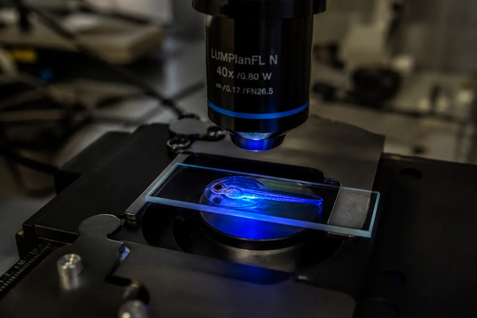
When you actually want to watch a growth cone navigate a living, intact circuit in real time, you step up to optogenetic axon guidance. The 2021 protocol for focal light activation of photoactivatable Rac1 (PA-Rac1) in embryonic zebrafish is the cleanest version of this I've seen — you pulse blue light at a specific region of the axon, you get a controlled cytoskeletal rearrangement, and you can steer growth trajectories through or even past endogenous repulsive barriers. You're no longer inferring what the cone would do; you're driving it and watching the system respond.
This is where the habenulo-interpeduncular pathway becomes your gold-standard in vivo assay. It's a beautiful system: dorsal habenular (dHb) axons reach their midbrain target, the interpeduncular nucleus (IPN), between 2 and 3 days post-fertilization, while the ventral habenular (vHb) nuclei aren't even fully formed until 4 dpf and only project later. That gives you two populations, two timelines, and a single target — perfect for comparing how the same guidance environment shapes two distinct axon cohorts. With PA-Rac1, you can experimentally force a dHb axon to take a vHb-like trajectory, or stall it before the IPN, and ask what happens to downstream circuit assembly. You can't ask that question in a stripe assay. The tissue tells you things the dish can't.
What makes this approach especially powerful for assay selection is the built-in internal comparison. You're not just asking "does the axon get there?" — you're asking "does it get there the right way, at the right time, using the right molecular logic?" Two populations converging on the same target through the same tissue, but on different developmental schedules, give you a level of experimental leverage that no purely in vitro system can replicate. The dHb axons arriving at the IPN by 72 hpf while vHb projections are still forming let you dissect timing-dependent guidance mechanisms without needing to engineer artificial conditions.
One caveat worth naming out loud: optogenetic steering doesn't fully bypass all endogenous guidance cues, and there's a real risk of off-target misrouting if your photoactivation region is too broad or your light dose is too high. Treat PA-Rac1 as a perturbation you calibrate against your control scaffold geometry at 24 hpf, not as a magic wand that overrides the brain.
The Hb-IPN pathway gives you a built-in internal control — same target, different developmental timing, same prep window.
Compartmentalized Analysis: Microfluidic Models for Adult Regeneration
Now flip the question. You're not studying embryonic development — you're studying regeneration in an adult. Maybe you're working with retinal ganglion cells (RGCs) after optic nerve injury and you want to watch dendritic remodeling and mitochondrial dynamics in real time in a single identified neuron. That's where the 2023 microfluidic adult zebrafish retinal model earns its place on the bench.
The microfluidic device compartments isolate the soma from the axon, fluidically separating cell bodies and axonal compartments so you can treat, image, and biochemically sample each side independently. You load an adult RGC, you watch its dendritic arbor remodel after injury, and you can pull mitochondrial dynamics data on a single-neuron basis. No culture-wide averages, no population noise masking the response of the cell you actually care about. That's a fundamentally different signal than anything an in vitro stripe prep can give you, and it's the only assay in this lineup that lets you ask adult-specific questions at single-cell resolution.
There's a practical reason this matters beyond the obvious appeal of single-cell data. Adult neurons behave differently than embryonic ones — their growth cone morphology, their cytoskeletal dynamics, their metabolic demands, and their responsiveness to guidance cues all shift with maturation. An assay designed for adult cells respects those differences rather than trying to force adult biology into an embryonic framework. If you've been running embryonic stripe assays and then wondering why your adult RGC data looks inconsistent, the mismatch between assay and cell type may be the problem.
What it cannot do is model embryonic spinal cord development directly. Adult RGCs and embryonic spinal neurons are not interchangeable systems, and the protocol modifications you'd need to swap one for the other are not trivial. If your lab is pivoting from regeneration to development, expect to rebuild at least half the prep and re-validate your compartment geometry before you trust any cross-system comparison.
Selecting the Right Assay for Your Question
So — which assay, when? Here's how I'd frame it for a trainee coming to me with a new project:
| Your question | Best fit assay | Why it fits | Time to first clean dataset |
|---|---|---|---|
| Does cue X repel growth cone Y on a flat substrate? | Stripe assay + primary culture | Binary substrate preference, high spatial control, 10–12 carpets in 4–5 h | ~1–2 weeks |
| How does a growth cone respond to a graded, diffusible cue? | Growth cone turning assay | Localized pulse, single-cell resolution, but gradient geometry is fragile | ~2–3 weeks |
| How does an axon navigate a living circuit in real time? | Optogenetic (PA-Rac1) in embryonic zebrafish Hb-IPN | Intact tissue, dHb/vHb comparison built in, 24 hpf scaffold ready | ~3–4 weeks |
| How does an adult neuron remodel during regeneration? | Microfluidic adult zebrafish RGC model | Compartmentalized soma/axon, single-neuron dynamics | ~4–6 weeks |
If you're chasing a mechanistic receptor–ligand question, start with stripes. If you're chasing how a circuit wires up in a living brain, jump to Hb-IPN. If you're chasing regeneration in a single identified adult neuron, microfluidics. The trap is trying to make one assay answer all three — and ending up with a beautiful prep that answers none of them cleanly.
A few practical rules of thumb I want you to internalize. First, never run a manipulation assay before you've characterized your baseline kinetics — know your extension rate, your substrate responsiveness, your control turning angles for your specific neuron type and prep. If your controls aren't behaving, your experiment isn't behaving either — you're just measuring noise with extra steps. Second, treat gradient geometry as a first-class variable in any turning or diffusion-based assay; the cue concentration at the cone is not the concentration in your pipette. Third, when you're working in an intact preparation, lean on the endogenous developmental schedule — the timing of dHb versus vHb arrival at the IPN, the 24 hpf scaffold — as built-in controls you didn't have to engineer. And fourth, when you read published rates and benchmarks from other labs, treat them as orientation points for your own system rather than universal truths — your neuron type, your substrate, your buffer conditions, and your animal husbandry all contribute to a baseline that's yours to characterize, not to match.
Here's the move I want you to make before you set up your next experiment. Write down, in one sentence, what question you're actually asking. Then check whether the assay you're about to run can answer that question, or whether it's just the assay that's most familiar to you. If those two answers diverge, that's your bottleneck — not your pipetting, not your reagent quality, not your imaging. The prep is downstream of the question, never the other way around.
Run the prep that fits the question. Get your baseline kinetics clean before you trust your manipulation. Know your numbers for your system, know what published ranges look like for context, and use that knowledge to catch problems early — not to impose a universal pass/fail grade on biology that's more variable than any single benchmark can capture.
Now go fix the noise in your assay — you've got good biology waiting on the other side.