Engineered Brain Organoids Reveal How the Cortex Maps Its Spatial Identity
By guiding brain organoids toward distinct front-and-back identities, researchers at UC Irvine have pulled back the curtain on a question that has long hovered at the edge of developmental…
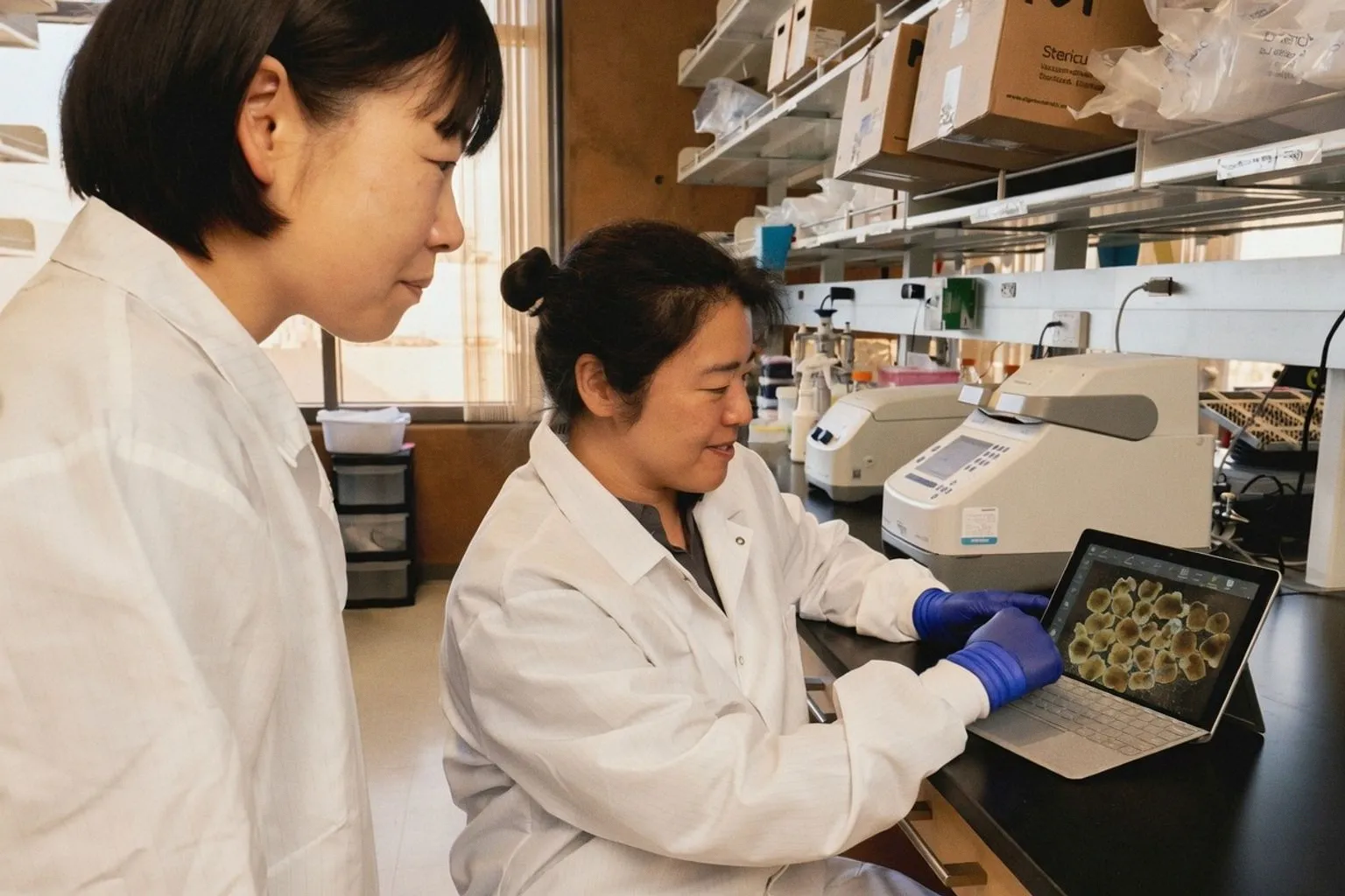
By guiding brain organoids toward distinct front-and-back identities, researchers at UC Irvine have pulled back the curtain on a question that has long hovered at the edge of developmental neuroscience: how does the cortex learn where it is?
The study, published this week in Cell Stem Cell, introduces lab-grown human neocortical tissue with engineered regional specificity, mimicking the spatial choreography of arealization, the chemical process by which the developing brain maps itself into functional zones. The advance reframes brain organoids from curious approximations into spatially resolved models, opening a clearer window onto the earliest acts of neural positioning.
A coordinate system written in chemistry
The cerebral cortex arrives in the world already patterned. Even before birth, gradients of signaling molecules help parcel the outer sheet into regions destined for vision, movement, language, and higher cognition. Conventional organoids tend to scramble this map, producing patchworks of mixed identities rather than coherent zones.
The UC Irvine team, working through the Sue & Bill Gross Stem Cell Research Center, exposed developing organoids to carefully selected morphogen signals early in their growth, steering some toward frontal cortex character and others toward posterior features. Single-cell profiling confirmed that the resulting tissues resembled actual human cortical development in their regional distinctions. Ordinary brain organoids replicate many features of developing tissue, but they usually miss this step; each one ends up with a random patchwork rather than a clear front or back.
Why position inside the cell matters
A companion line of inquiry, reported last month in Nature by researchers at UCSF, adds another spatial dimension to the same question. As neural stem cells mature into neurons, developmental genes shift from the nuclear periphery toward nuclear speckles, structures dense with RNA-processing machinery. The repositioning correlates with stronger gene expression; among genes that detached from the nuclear lamina, roughly 41 percent migrated to speckles, including SATB2, MEF2C, and SYT1, all linked to neurodevelopmental conditions.
The implication is structural: the location of a gene inside the nucleus may help determine whether it stays silent or switches on, complementing the chemical tags that have long dominated epigenetic study. Position, it seems, is a form of regulation in its own right.
What to watch in the lab
For researchers working with organoid or animal models, the practical takeaway is architectural. When designing experiments that ask why one cortical region behaves differently from another, build the spatial axis into the model from the first passage, not as a downstream annotation. Chemical gradients, timing, and substrate geometry all shape regional identity; treating the tissue as homogeneous will flatten the very differences that matter.
And when interpreting gene expression data, consider that a gene's address, its proximity to the nuclear lamina or to speckles, may weigh as heavily as its chemical modifications. Both lines of work converge on a single principle: in the developing brain, where something sits shapes what it becomes.