Neural Flexibility: How Brain Circuits Reuse Populations for Sensory and Motor Tasks
The MIT group behind it, led by postdoctoral researcher Yuma Osako, has produced what outlets like Medical Xpress and Rediff are calling the first evidence that prefrontal cortex neurons swap roles…
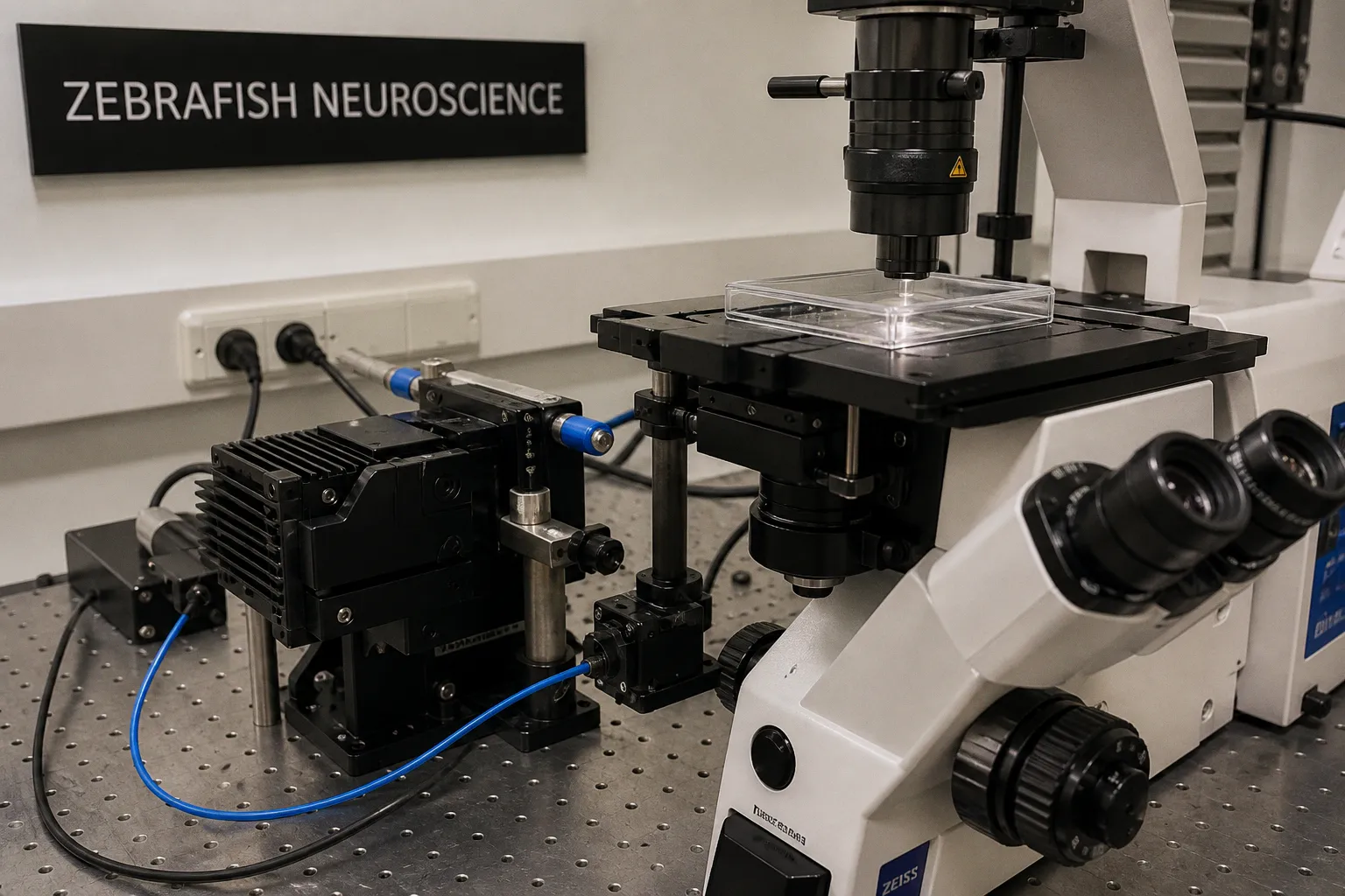
If you've ever stared at your two-photon traces wondering why the same cell pops off during a sensory stimulus AND during a motor decision, congratulations - your data might not be noisy. It might be telling you exactly what a new Nature Neuroscience paper just told the rest of the field: the brain reuses the same neural populations for different jobs. The MIT group behind it, led by postdoctoral researcher Yuma Osako, has produced what outlets like Medical Xpress and Rediff are calling the first evidence that prefrontal cortex neurons swap roles between holding a sensory memory and storing an action plan in working memory.
The reusable module idea, finally with receipts
We've talked about "mixed selectivity" and "flexible coding" at lab meetings for years, but this is the first time anyone's pinned it down with a prep rigorous enough to make the claim without hedging: the same subset of neurons can hold both an action and a sensory stimulus in working memory at different moments. Osako's team trained mice on a two-tone matching task and recorded electrical activity across prefrontal and parietal cortex. The parietal neurons stuck to one job - tracking the tone. The prefrontal ones flipped: during the delay after the first tone, they were holding the sensory memory; during the delay before the response, they were carrying the action plan.
That kind of role-switching within a single trial is the thing your own prep might be seeing, but you've probably been filing it under "artifact" or "movement-related bleed-through." The MIT data argue it's a feature, not noise.
What to actually do at the bench
So let's talk about how to catch this in your own animals. If you're running a similar delayed-response paradigm in zebrafish larvae - and many of us are, given how clean the optical access is - here is where I would push you to look twice:
- Audit your trial structure. Are you pulling apart the delay windows in your analysis? Osako's group looked at two distinct epochs, between tone 1 and tone 2, and between tone 2 and the decision. If you're averaging across the whole ITI, you are smearing the very signal they just isolated.
- Re-examine your "movement neurons." The cells that fire before the swim turn or the saccade are usually the first ones we mark for exclusion. Ask whether they are carrying a plan rather than executing a movement - you will need tighter alignment to stimulus and response onset to tell.
- Push for population-level analyses. Single-cell decoding will keep telling you "this neuron responds to X." Dimensionality reduction or state-space approaches are where the role-switching becomes visible, which is exactly what the MIT team used to identify their reusable clusters.
Why this reframes the synaptic switch question
Here is the through-line: the framing you will see in Medical Xpress as a "reversible synaptic switch" is not metaphor. If the same physical neurons can carry different computational content depending on the moment and the task demand, then plasticity at those synapses is not about rewriting a fixed circuit - it is about toggling what the circuit does. For anyone doing long-term imaging of the same identified cells across developmental stages or learning epochs, this validates the frustration you have probably had with a strict "labeled line" interpretation. Your prep's job is to build the conditions where that toggling can be seen cleanly, and the rest of us will be watching to see whether zebrafish circuits show the same reusable logic - my money says yes, but we will need the data.