Reprogramming Reactive Astrocytes into Functional Neurons to Repair Spinal Cord Injuries
According to Medical Xpress, a joint team led by C.
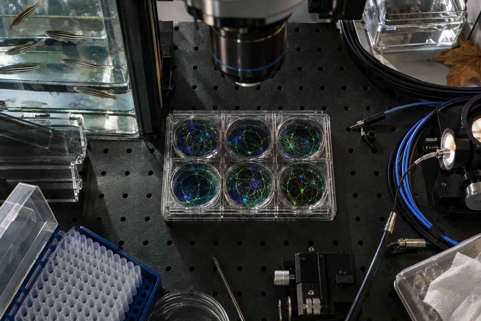
Justin Lee at the Institute for Basic Science and Ha Yoon at Yonsei University College of Medicine has built a genetic system that converts scar-forming astrocytes into functional neurons at spinal cord injury sites, with measurable motor recovery in mice and rats. The construct, named TRANsCre-DIONE, uses a two-signal molecular switch that gates a neuronal programming gene only in the reactive astrocyte population, sidestepping the targeting errors that stalled earlier conversion strategies. The findings, published in Experimental & Molecular Medicine, read less like a therapy and more like a clean methodological template: here is how you repurpose local glia to rebuild broken circuits, here is how you verify it works.
The targeting problem
Reactive astrocytes are an obvious substrate for repair. They populate the lesion, they survive, and they sit exactly where lost neurons used to fire. The bottleneck has always been selectivity. Prior astrocyte-to-neuron conversion work leaned on the GFAP promoter alone — but GFAP is active in healthy astrocytes and in some neural progenitors, and its expression tends to decline once a cell begins adopting neuronal identity. The very switch driving reprogramming can flick off mid-process.
TRANsCre-DIONE isolates the reactive population with an AND-gate logic. It pairs GFAP regulatory elements with elements tied to Lcn2, an inflammation-linked protein elevated at the injury site. Both signals must be present in the same cell before the construct activates Neurog2, a transcription factor that pushes cells toward neuronal fate. A second regulatory element then sustains Neurog2 expression, so conversion persists even after the astrocyte's original molecular signature fades. Clean input, sustained output.
What the data show
The construct was delivered near spinal cord lesions in mice and rats two weeks after injury. In treated mouse cords, 87% of labeled cells carried a neuronal marker, and subsets expressed motor-neuron markers Isl1 and ChAT. Electrophysiological recordings confirmed the new cells could fire action potentials and receive synaptic input — evidence of integration into local circuitry rather than molecular mimicry.
On a nine-point locomotor scale, treated mice reached an average of 3.88 at eight weeks, while controls stayed below 1. Treated rats showed the same direction of effect. The team followed up with functional tests to confirm the new neurons were actually driving the recovery, not just accompanying it.
What to calibrate next
For practitioners tracking in vivo reprogramming, the open parameters are familiar: how long Neurog2 expression must be sustained to stabilize converted neurons, whether off-target conversion occurs in non-lesion tissue, and whether the new cells wire into correct long-range loops rather than local shortcuts. The motor gains are encouraging, but read as a proof-of-concept signal, not a clinical benchmark — the approach has not been tested as a treatment in humans. Human translation will hinge on whether reactive astrocytes in human injuries carry the same dual GFAP/Lcn2 signature, and whether Neurog2 induction in that context produces comparable functional output.