High-Speed 3D Imaging System Maps Seizure Propagation in Zebrafish Brains
If you've ever stared at a 2D seizure trace in your larval zebrafish prep and thought, "I'm sure this thing is happening somewhere I can't see," you're not alone.
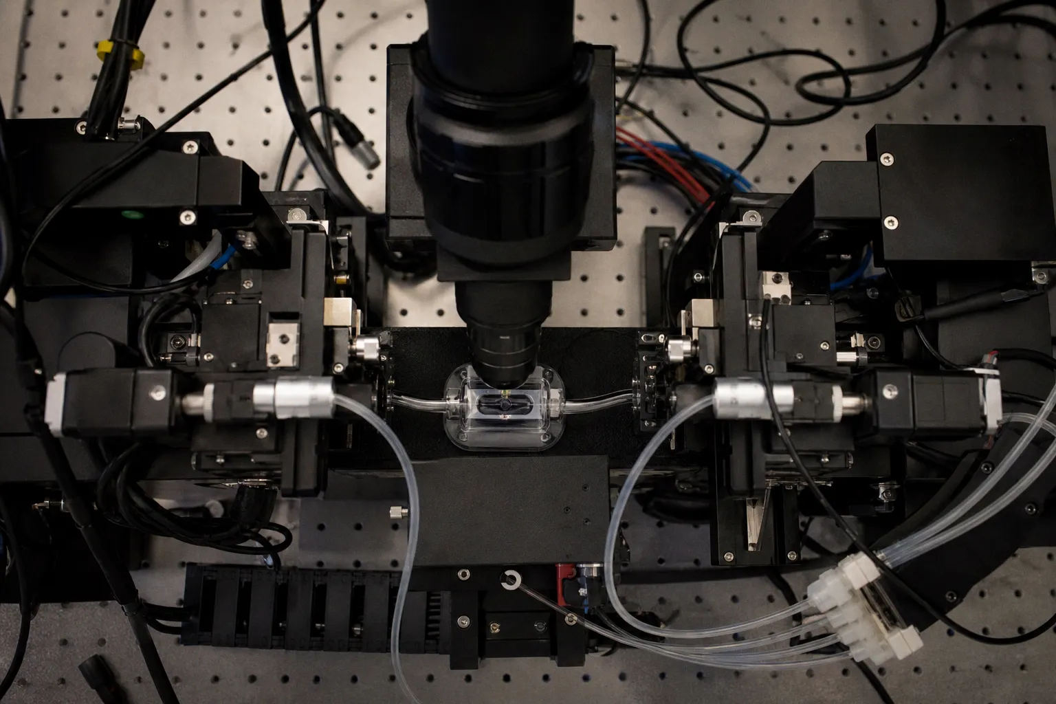
A team at the University of Georgia just built the kind of rig that would have saved you a year of guesswork: a high-speed light-sheet system paired with adaptive optics that finally films a full seizure rolling through the brain in 3D, start to finish.
What's actually new at the bench
The setup, described in work led by Peter Kner's group, combines two tricks zebrafish labs have wanted for a long time. First, light-sheet illumination — a thin optical slice instead of a cone of excitation — keeps phototoxicity and background noise low enough that you can image a behaving larva at speed. Second, and this is the part that matters when you're chasing millisecond-scale events, adaptive optics borrowed straight from astronomy corrects the blur introduced by the tissue itself. The light bends on its way in, and adaptive optics bends it back. The result is the clean signal you've been trying to coax out of your own scope for months.
In the published seizure recordings, the electrical wave originates toward the back of the brain and sweeps forward into the optic tecta — the midbrain hub handling visual processing and eye movement — before subsiding over several seconds. That propagation path is exactly the kind of thing 2D imaging tends to hide, because a single plane will always make you wonder whether you caught the whole event or just a slice of it.
What this means for your zebrafish prep
Let's be honest about why this is exciting and why you shouldn't go tearing down your rig tomorrow. The Kner lab's system is engineered for whole-brain volumetric capture at seizure-relevant timescales, and adaptive optics is what makes the difference between "I can see something is happening" and "I can tell you where, in three dimensions, at this exact frame." For anyone running PTZ or pentylenetetrazol-kindling assays, optogenetic induction, or genetic epilepsy models in zebrafish, the practical question is no longer whether 3D propagation exists — it's whether you can resolve the sub-regions where it begins.
What I'd watch for next: whether the optical recipe travels outside UGA. Adaptive optics on a light-sheet isn't plug-and-play yet, but the principle — correct for tissue-induced wavefront distortion rather than just averaging harder — is the direction the whole field is moving. If you're designing a new imaging rig or writing a methods chapter, cite this one. It sets the bar.
A small honest caveat
You're not going to replicate the full setup this quarter, and you don't need to. The right takeaway is methodological: if your seizure phenotype data has felt suspiciously clean and suspiciously flat, the problem might not be your fish. It might be that you're watching in 2D. Start with what you have — a faster acquisition protocol, a thinner optical section, even a carefully registered z-stack — and you may find that the propagation story you've been missing was always there, just outside your plane of focus. Keep pushing.