Zebrafish light-sheet imaging: 4 phototoxicity fixes
A larval zebrafish at 48 hours post-fertilization presents an imaging scientist with a quiet paradox: the photons required to resolve its nascent neurons with subcellular clarity are also capable of changing the physiology under observation.
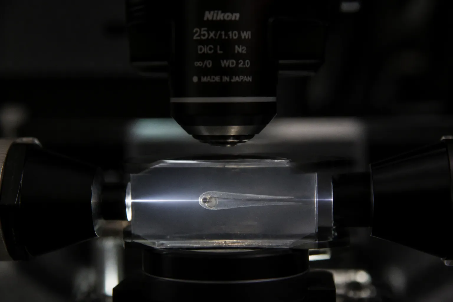
Light-sheet fluorescence microscopy was designed to reduce that conflict. By confining excitation to a thin optical plane rather than sweeping a focused point across an entire volume, LSFM can substantially reduce the light dose delivered to the specimen. The approach was recognized as Nature Methods’ Technique of the Year in 2014, in part because of this combination of optical sectioning and comparatively gentle illumination.
But “minimizes” is not the same as “eliminates.” A light sheet can still produce photobleaching, local heating, and reactive oxygen species, particularly when a fluorescent specimen is illuminated repeatedly over a long experiment. The biological consequences are not fixed in advance: they depend on wavelength, power, exposure time, indicator chemistry, oxygenation, mounting, acquisition geometry, and the sensitivity of the developmental or neural readout. Calcium dynamics, developmental progression, and behavior can all become harder to interpret if the imaging protocol itself is not treated as an experimental variable.
The fix is rarely a single upgrade. It is a layered discipline applied to how photons enter, travel through, and leave a living brain. Four strategies are especially useful: changing the geometry of the beam, moving excitation toward the near-infrared, breaking continuous illumination into controlled intervals, and using transmitted-light tomography when fluorescence is not necessary for the question.
Thin the sheet: lattice and Bessel beam geometry
The first decision any light-sheet operator makes is the shape of the illumination plane. A conventional Gaussian light sheet, formed by focusing a beam through an illumination objective, is thinnest around its waist and gradually expands away from it. That is a fundamental limitation of the beam rather than an alignment error. Across a large field of view, different parts of the specimen may therefore experience different combinations of sheet thickness, intensity, and optical sectioning quality.
For a larval zebrafish whose brain spans only a few hundred micrometers, this non-uniformity matters. A region close to the beam waist may be sectioned efficiently, while a peripheral or deeper region may require more excitation to produce a comparable signal. Increasing power can rescue brightness, but it also raises the dose delivered to tissue that may already be close to the required signal-to-noise threshold.
Lattice light-sheet microscopy addresses the problem by replacing the smooth Gaussian profile with a structured optical lattice. The excitation plane is assembled from the interference of multiple coherent beams, creating a thin and extended sheet. The aim is not simply to make the sheet narrow at one point, but to maintain useful optical sectioning over a broader area while limiting unnecessary illumination outside the active plane.
Bessel beam illumination takes a different route. A Bessel-like beam has a narrow central core that remains comparatively stable over a longer axial range than a conventional Gaussian waist. This can be valuable when the field of view is large or when the specimen’s geometry makes it difficult to keep every relevant structure near the ideal focus. The trade-off is that Bessel beams generate side lobes. Those additional intensity bands can excite fluorophores outside the central core, so the optical design and the detection strategy have to be considered together.
Neither method should be described as automatically gentle. A thinner or more uniform sheet can reduce the amount of excitation needed for a given image quality, but the actual benefit depends on how the system is aligned and operated. The same beam geometry can produce very different biological outcomes if the operator compensates for poor transmission by increasing power, extending exposure, or repeating acquisitions unnecessarily.
A thinner illumination plane is not a guarantee of harmless imaging. It is a better starting point for deciding where the photons should go.
For zebrafish laboratories moving from standard Gaussian SPIM, the practical question is whether the gain in dose efficiency justifies the added optical complexity. Lattice systems require careful calibration of the interference pattern and stable control of the illumination path. Bessel implementations require attention to side-lobe excitation, beam shaping, and the relationship between illumination and detection numerical apertures.
A useful comparison begins with the experiment rather than the name of the beam:
- Large field of view: a structured or extended beam may distribute excitation more evenly than a tightly focused Gaussian sheet.
- Deep or irregular anatomy: a Bessel-like profile can preserve a narrow central region over a longer distance, but side-lobe exposure must be managed.
- Long developmental series: reducing the required intensity per volume may be more valuable than achieving the thinnest possible sheet at a single location.
- High-resolution cellular imaging: the limiting factor may be the fluorophore and detector rather than beam geometry alone.
The correct metric is not whether the instrument uses lattice or Bessel illumination. It is whether the same biological information can be acquired with less excitation, fewer redundant planes, or fewer repeated passes through the same tissue.
Shift the wavelength: near-infrared two-photon excitation
Visible-wavelength light sheets can work exceptionally well in transparent early larvae, especially when the reporter is bright and the structure of interest is close to the illumination surface. As the animal grows, however, scattering and absorption become more consequential. Deeper structures such as the hypothalamus, hindbrain, and ventral brain may be difficult to image with the same efficiency as superficial regions.
Near-infrared excitation offers a different optical compromise. In two-photon microscopy, the fluorophore absorbs two lower-energy photons in a nonlinear event. Excitation is concentrated where the photon density is highest, and near-infrared wavelengths generally travel through tissue more effectively than shorter visible wavelengths. This can improve access to deeper structures and reduce out-of-focus excitation, although it does not remove phototoxicity from the experiment.
The distinction between peak and average power is central. Two-photon excitation relies on high instantaneous intensity, usually delivered in short pulses, while the average power at the specimen determines much of the longer-term thermal and photochemical burden. A protocol may therefore have high peak pulse power without producing the same average exposure as a continuously illuminated single-photon setup. That comparison is meaningful only when pulse duration, repetition rate, dwell time, scanning pattern, wavelength, and specimen response are considered together.
Power values reported for two-photon systems should be treated as experimental reference points, not universal safety limits. The threshold at which a larva begins to show physiological or developmental effects depends on the optical path, the fluorophore, the mounting geometry, the rate of heat removal, and the endpoint used to detect damage. A setting that is acceptable for a short structural acquisition may be inappropriate for a long calcium-imaging session.
In larval zebrafish, implementation commonly pairs a tunable femtosecond laser with a low-NA illumination objective and a high-NA water-immersion detection objective. Ti:sapphire excitation around the range used for GFP- and GCaMP-class reporters is a familiar starting point, but the optimal wavelength is not simply the one that produces the brightest image. It is the one that provides sufficient signal while preserving the biological state of the specimen.
Mounting and environmental control become particularly important when average power is high or the imaging session is long. Immobilization in low-melting-point agarose is common, with the gel surrounded by physiological embryo medium. The sample chamber must support stable temperature, adequate oxygen exchange, and enough circulation to prevent local heating. A larva can remain optically stable while its environment drifts physiologically, so image quality alone is not a sufficient indicator that the protocol is benign.
Several checks help keep near-infrared two-photon imaging honest:
1. Separate optical performance from biological tolerance. A deeper, brighter volume is not automatically a better volume if the preparation shows altered activity or delayed development.
2. Measure the specimen plane, not just the laser display. Losses in the objective, scanner, and illumination path can make nominal settings misleading.
3. Compare matched biological endpoints. Monitor fluorescence stability alongside movement, heart activity where relevant, developmental progression, or neural baseline.
4. Avoid treating a single threshold as portable. A power limit established for one reporter, objective, or chamber should not be transferred unchanged to another preparation.
Near-infrared two-photon excitation is best understood as a way to redistribute the optical burden. It can improve depth and confine excitation, but the energy still reaches a living organism. The protocol must still account for heat, oxygen chemistry, fluorophore behavior, and the cumulative effect of repeated illumination.
Edit the time: intermittent and stroboscopic illumination
Spatial optimization eventually reaches a ceiling. If the experiment requires repeated volumetric imaging, the specimen will still be exposed many times even when each individual light sheet is carefully shaped. The remaining variable is temporal: how often the larva is illuminated, how long each exposure lasts, and whether every acquired plane contributes meaningfully to the biological question.
Intermittent illumination, also described as strobed or pulsed acquisition, replaces continuous exposure with synchronized bursts. The camera, scanner, and illumination source are coordinated so that the specimen is lit only during the portion of the acquisition that carries useful information. Between bursts, the excitation is off. This does not make the experiment risk-free, but it can reduce cumulative exposure and limit the time available for heat and photochemical products to build up.
The protocol has to follow the dynamics of the biology. Calcium indicators such as GCaMP report signals that unfold over a characteristic timescale rather than requiring uninterrupted illumination at every moment. If the scientific question concerns population-level activity, event timing, or slower developmental changes, continuous excitation may provide little additional information. If the experiment aims to resolve very fast transients, aggressive temporal sparsening could erase the signal of interest.
There is no universal volumetric rate or dose-reduction factor that applies to every zebrafish preparation. A useful schedule depends on the indicator’s kinetics, the expected event frequency, the number of planes, the camera’s readout, the illumination duty cycle, and the reconstruction method. The relevant comparison is between two validated protocols that deliver comparable information—not between an abstract “continuous” setting and a nominally “intermittent” one.
A practical stroboscopic design usually begins by defining the minimum temporal resolution needed for the biological event. From there, the operator can ask:
- Can some planes be sampled less frequently than others?
- Does every volume need the same exposure time?
- Can illumination be synchronized to the camera’s active acquisition window?
- Can a lower-intensity preview identify periods that deserve denser sampling?
- Is the reported activity stable when the interval between acquisitions changes?
The last question is especially important. Intermittent illumination can alter the apparent baseline if the reporter or tissue does not recover in the same way between exposures. Photobleaching may be slower, but the signal can still decline. Conversely, a protocol that reduces exposure too aggressively may produce sparse data that are later amplified by denoising or motion correction, creating the impression of activity that the raw signal does not support.
Thermal management benefits from the same discipline. Continuous laser exposure can create local temperature gradients, particularly around the illumination objective, chamber walls, or poorly mixed mounting medium. Intervals without excitation give the system an opportunity to dissipate heat, but the recovery time is determined by the chamber and environment. It should be checked rather than assumed.
The best stroboscopic protocol is not the one with the darkest gaps. It is the one that preserves the biological timescale while removing illumination that the analysis never needed.
For long developmental imaging, this approach is often more flexible than simply lowering laser power. Lowering power may reduce signal below the detector’s useful range; shortening or spacing exposures can preserve per-frame quality while reducing the total number of illuminated seconds. The two adjustments can also be combined, provided the resulting signal is validated against an appropriate control.
Borrow the context: bright-field optical tomography
Fluorescence is indispensable when the experiment requires molecular specificity or activity reporters. It is not indispensable for every structural question. When the goal is to track gross anatomy, tissue boundaries, overall morphology, or specimen drift, transmitted-light imaging can provide valuable context without repeatedly exciting a fluorescent molecule.
Bright-field optical tomography reconstructs three-dimensional structural information from transmitted white-light images acquired at multiple angles. The specimen is still illuminated: this is not a no-light method. Its advantage is that it avoids fluorescence excitation and the associated repeated excitation of fluorophores. Absorption and refraction contribute to the recorded contrast, allowing a label-free structural reference to be generated alongside or between fluorescence acquisitions.
That distinction matters when describing phototoxicity. Transmitted light can still contribute to heating or other optical stress, depending on intensity, wavelength, exposure duration, and the geometry of the preparation. The appropriate claim is therefore reduced fluorescence-related burden or a lower-dose structural modality—not guaranteed absence of phototoxic effects.
Optical tomography also does not replace functional fluorescence. A refractive-index map cannot identify the activity of a GCaMP-expressing neuron or distinguish one molecular reporter from another. Its value lies in reducing the number of times fluorescence must be used for information that is fundamentally anatomical.
In a multimodal workflow, tomography can be placed at strategic intervals:
- a structural volume before the functional run establishes the initial anatomy;
- selected transmitted-light acquisitions during the session can help identify drift or gross morphological change;
- a final structural volume provides a reference for registration and for checking whether the specimen retained the expected shape.
The frequency should follow the purpose of the structural check. If the anatomy is stable and the experiment is focused on neural activity, repeated fluorescence volumes may be unnecessary. If the study follows rapid morphogenesis, a more frequent low-dose structural record may be justified.
Registration between transmitted-light and fluorescence data requires care. The two modalities do not report the same contrast, and a boundary visible in bright-field may not correspond to the edge of a fluorescent structure. Fiducial features, stable anatomical landmarks, and consistent mounting improve alignment. The structural channel should support the functional interpretation, not be treated as a perfect ground truth.
A compact comparison
| Strategy | Primary mechanism | Where it helps | Main trade-off |
|---|---|---|---|
| Lattice or Bessel illumination | A thinner or more extended structured excitation profile | More even illumination and potentially lower excitation per useful voxel | Alignment complexity; Bessel side-lobe control |
| Near-infrared two-photon excitation | Longer-wavelength nonlinear excitation with localized signal generation | Deeper access and reduced out-of-focus excitation in suitable preparations | High instantaneous intensity; heat and oxygenation still require control |
| Intermittent illumination | Synchronized light bursts matched to the biological timescale | Lower cumulative exposure and better control of thermal load | Requires careful timing, synchronization, and validation of temporal fidelity |
| Bright-field optical tomography | Transmitted-light structural contrast without fluorescence excitation | Anatomical context, drift tracking, and label-free reference volumes | Still uses light and provides no molecular specificity |
The table is a design aid, not a ranking. These strategies address different sources of burden. Beam geometry changes where light is deposited. Wavelength changes how it propagates and how excitation is confined. Timing changes how long the specimen is exposed. Tomography changes which questions need fluorescence at all.
The editorial eye applied to photons
Reducing phototoxicity in larval zebrafish light-sheet imaging is rarely a matter of purchasing one more optical component. It is a sustained editorial discipline: deciding which photons carry information, which are redundant, and which are being used merely because the instrument is already running.
The four strategies above operate on different physical axes—geometry, wavelength, time, and modality. Their benefits are cumulative, but so are their assumptions. A lattice sheet does not compensate for unnecessary repeated volumes. Near-infrared excitation does not make high average power harmless. Stroboscopic acquisition does not preserve a signal that is sampled too slowly. Transmitted-light tomography does not provide molecular specificity simply because it produces a three-dimensional image.
A robust protocol therefore treats phototoxicity as something to measure alongside the primary result. Fluorescence intensity, bleaching, motion, survival, developmental progression, spontaneous activity, and behavioral output can each reveal a different kind of disturbance. The most useful control is not always a separate untreated animal. It may be a matched imaging condition with altered duty cycle, reduced volume rate, lower excitation, or a structural modality substituted for part of the fluorescent acquisition.
Functional fluorescence imaging carries phototoxicity risk; whether that risk matters depends on dose, timing, tissue, reporter, and the endpoint being measured.
This is also why “safe” settings should be described with restraint. A protocol that preserves morphology may still affect neural activity. One that leaves calcium transients apparently intact may alter later behavior or development. The absence of visible damage is evidence, but it is not proof that the optical perturbation is irrelevant to every downstream measurement.
The practical principle is simple: treat photon delivery as a budget. Match the dose to the question rather than to the convenience of continuous acquisition. Use the thinnest and most efficient illumination that produces the required signal, move to a wavelength and modality that suit the depth, and turn the light off whenever the experiment does not need it.
A larva that remains physiologically credible is more valuable than a brighter volume. In developmental neurobiology, the goal is not merely to see the brain for longer. It is to preserve the conditions under which the brain can continue to reveal what it is doing.