FlatMux Platform Overcomes Two-Photon Imaging Limits for Zebrafish Neurobiology
A new platform called FlatMux, described in Nature Methods by Alipasha Vaziri's group at Rockefeller and reported by The Transmitter, is built to win that fight — letting you image genetically…
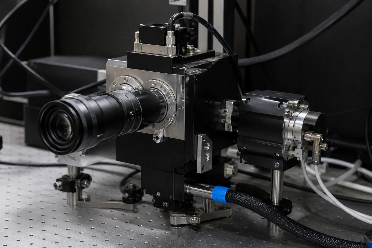
Anyone who has tried to pull a clean voltage trace from a behaving larva knows the old two-photon trade-off. A new platform called FlatMux, described in Nature Methods by Alipasha Vaziri's group at Rockefeller and reported by The Transmitter, is built to win that fight — letting you image genetically encoded voltage indicators across wider fields, deeper tissue and even multiple depths simultaneously. For zebrafish labs running circuit-level physiology, that combination is exactly what we have been begging our scopes to do for years.
The old voltage ceiling
Calcium indicators have been our workhorse for more than a decade — they flash when calcium floods into a firing neuron, and they get the job done most days. But calcium is a proxy, and a slow one; it smears the millisecond-scale timing that real voltage events live in. GEVIs watch the membrane potential directly, which sounds like a dream until you remember what they actually give you in practice: signals that last only around a millisecond, sensors that sit in membranes fighting your objective, and coverage that usually stops at about 50 by 250 micrometers over maybe ten neurons. If your zebrafish prep has even a moderately active tectum or habenula circuit, you have already lost half the population before you press record. Add the photobleaching risk and the temptation to crank the laser, and you can see why most of us have stuck with the calcium workflow we know is good enough.
What FlatMux actually changes
Think of the old system as a juggling act between three knobs we never stop fighting: energy, time and space. A point-scanning two-photon has to fire at roughly a thousand frames a second or faster to catch a GEVI blip, and as Jerry Chen at Boston University puts it, "you have to walk this tightrope, where you put enough light into the brain to get signal out, without frying the brain." Push too hard and you bleach the sensor; back off and your signal disappears into noise. Vaziri's group tunes all three together — pulse energy, timing and pixel density — so you end up with essentially one pulse per pixel instead of wasted dwell time. The payoff is that you can sweep wider fields and trace activity through different cortical layers at the same time, which has typically demanded two separate runs and a lot of co-registration prayer.
Where to place your bet this quarter
Two practical moves to make at the bench this month. First, FlatMux is published as a reproducible optical design, so reach out to the Vaziri lab early if you want to co-build or co-test a copy — most of the real bottlenecks only show up at that handoff step, not in the paper figures. Second, and this is the part our community often skips, sanity-check which GEVI lines actually express cleanly in zebrafish. Most current GEVIs were validated in mouse cortex, and membrane trafficking plus brightness can shift dramatically in fish. Run a small side-by-side pilot against your current calcium prep on the same neurons before you commit to a rig rebuild. Voltage imaging at depth is finally within reach — let's make sure our indicator and prep catch up before we buy in.