Calcium imaging systems for zebrafish: choosing the right setup
The most expensive mistake in zebrafish calcium imaging is not buying the wrong objective. It is building a system that produces beautiful movies while quietly disturbing the biology you meant to measure.
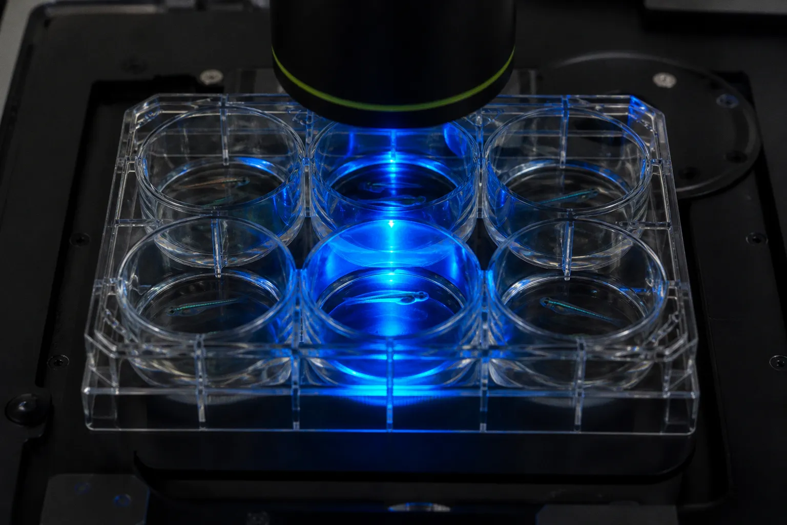
This happens most often during visuomotor experiments. A conventional one-photon light-sheet setup may use 488 nm illumination, which sits within the sensitive range of the zebrafish visual system. In a transparent larva, that light can reach the retina, excite photoreceptors, and become part of the stimulus. Your apparently clean neural response may therefore contain a response to the microscope itself. The fish is not being difficult. Your optics have joined the experiment.
So when we talk about zebrafish calcium imaging system selection criteria, we need to start with the experiment, not the instrument brochure. Are you mapping activity across the whole larval brain? Following a fast sensorimotor circuit? Recording during visual stimulation? Working with an adult fish that must breathe and move? Each question changes the balance between spatial resolution, frame rate, penetration depth, wavelength, and behavioral freedom.
Let’s walk through the choice as we would at the bench: first define the signal you need, then identify the noise your setup might introduce, and only after that decide which microscopy architecture earns its space, budget, and maintenance burden.
Start with the biological question, not the microscope
A zebrafish larva is typically about 3 mm long and transparent enough for in vivo optical imaging across much of the brain. That combination is extraordinarily useful, but it can tempt us into thinking the imaging problem is automatically easy. It is not. Transparency gives you access; it does not guarantee that every photon carries useful information.
For whole-brain functional imaging, the central challenge is usually not whether you can detect fluorescence. It is whether you can detect changes in fluorescence across a sufficiently large volume, quickly enough, with enough spatial detail to separate nearby structures, while leaving the animal’s behavior and sensory environment intact.
A good prep begins with four decisions:
- How much of the brain must be recorded at once? A small region may tolerate a point-scanning system; whole-brain mapping usually benefits from parallelized acquisition.
- How fast does the neural event unfold? If the circuit produces rapid activity changes, a slow volumetric stack can turn a clean event into temporal soup.
- What sensory signals must remain natural? Visual experiments are particularly sensitive to unintended illumination, but the same principle applies to thermal, mechanical, and acoustic disturbances.
- How freely must the fish behave? Immobilized larvae and head-restrained adults are not interchangeable preparations, even when the calcium indicator and microscope are similar.
This is why there is no universal setup that fits every zebrafish neurobiology project. A system optimized for fast whole-brain imaging in a larva may be a poor choice for deep imaging in an adult forebrain, and a system that is excellent for structural detail may be unnecessarily restrictive for a simple activity map.
What the calcium signal is asking from your hardware
Calcium indicators report neural activity indirectly. The fluorescence change depends on the indicator, expression level, excitation conditions, detector sensitivity, and the kinetics of calcium handling in the cells you are recording. Hardware cannot repair a poor indicator choice, and an excellent indicator cannot rescue a system that samples too slowly or floods the preparation with irrelevant light.
When considering a calcium indicator choice for zebrafish, connect the indicator to the recording goal:
- For broad population activity, you may prioritize coverage and signal-to-noise across many neurons.
- For rapid circuit dynamics, temporal sampling becomes more important than squeezing out the last increment of spatial resolution.
- For behavior-linked imaging, stable registration matters because movement creates a signal that can easily masquerade as neural activity.
- For long recordings, phototoxicity and bleaching become practical limits rather than footnotes in the methods section.
The clean signal is the one that survives all of these constraints. A bright movie is not necessarily a good recording.
The best imaging system is not the one with the most impressive specification. It is the one that introduces the least biological confusion.
Light-sheet fluorescence microscopy: speed and coverage with a catch
Light-sheet fluorescence microscopy is a natural fit for larval zebrafish brain imaging because it illuminates a plane rather than the entire optical path through the specimen. That optical sectioning reduces unnecessary excitation outside the plane of interest, while parallelized image acquisition allows the system to capture large volumes quickly.
For whole-brain functional imaging, this matters enormously. Instead of collecting a single point and building the image one pixel at a time, light-sheet architectures can acquire information across a plane and move through the volume rapidly. The result is high-speed volumetric imaging over the brain, which is exactly what you want when the biological question concerns distributed neural activity rather than one isolated neuron.
But light-sheet microscopy is not automatically gentle, and it is not automatically behaviorally neutral. The illumination wavelength is part of the experiment. Conventional one-photon systems commonly use visible excitation such as 488 nm, and that wavelength lies within the sensitive region of the fish’s visible spectrum. During a visual stimulation experiment, the excitation light can activate retinal photoreceptors.
That creates a particularly irritating form of noise: the microscope becomes an unplanned visual stimulus. You may see a population response and assume that your moving grating, flash, or looming stimulus caused it, when some fraction of the response came from the illumination geometry. If the timing is consistent, the artifact can even look beautifully reproducible. Reproducible is not the same as valid.
When conventional one-photon light-sheet remains useful
This does not make one-photon light-sheet microscopy useless. Far from it. It remains a powerful option when:
- the experiment does not depend on naturalistic visual stimulation;
- the illumination geometry can be shielded or controlled adequately;
- you need rapid volumetric acquisition across the larval brain;
- the preparation is sufficiently transparent for the depth and field of view you require;
- the main priority is broad functional coverage rather than deep adult-brain imaging.
The mistake is treating the illumination wavelength as a minor technical detail. It belongs in the experimental design from day one, alongside the behavioral stimulus and the indicator.
If the fish is exposed to 488 nm during the recording, ask a blunt question: could the retina detect this? If the answer is yes, design controls that separate neural responses to the intended stimulus from responses caused by the imaging beam. Better still, consider whether another excitation architecture can remove the problem rather than forcing the analysis to compensate for it later.
The 488 nm problem: when your microscope becomes part of the stimulus
Visual stimulation experiments are especially unforgiving because zebrafish larvae are not passive transparent containers for your optics. Their visual system is active, sensitive, and tightly connected to the behaviors many researchers want to measure.
A conventional visible-light sheet can therefore introduce interference in several ways:
1. Direct retinal activation. Illumination reaches photoreceptors and produces a visual response independent of the intended stimulus.
2. Timing confusion. Imaging light may be pulsed, scanned, or synchronized with acquisition in a way that overlaps with stimulus presentation.
3. Spatial ambiguity. A light sheet crossing the preparation can expose regions outside the field you thought defined the visual input.
4. False confidence from clean traces. A stable artifact may look like a biological signal if the controls do not isolate illumination from stimulation.
5. Analysis contamination. Whole-brain activity maps can assign the response to the wrong circuit when the visual system has been activated by the imaging path.
The practical fix is not simply to subtract a baseline. Baseline correction can remove drift; it cannot reliably reconstruct an experiment in which the animal received an unintended sensory input.
Start with the simplest control: record the same preparation under the imaging illumination while removing or altering the intended visual stimulus. If the response remains, the microscope has contributed to the phenotype. Then vary the timing and intensity of illumination where possible. The goal is not to prove that the artifact is small. The goal is to know whether the artifact is present, where it appears, and whether it overlaps with the biology you care about.
This is also where experimental discipline saves money. Before ordering custom hardware, sketch the light path and list every wavelength reaching the fish. Note the timing of each source. Mark the retina, the brain region of interest, and any reflective surfaces that may redirect illumination. Five minutes of optical honesty can prevent weeks of elegant analysis built on a compromised preparation.
Two-photon light-sheet microscopy: solving the wavelength and speed trade-off
Two-photon light-sheet microscopy changes the problem by combining excitation confinement with infrared wavelengths. That combination can support non-invasive whole-brain functional recordings while avoiding unintended activation of photoreceptors by visible excitation light.
The benefit is not merely that infrared light sounds more sophisticated. The wavelength is biologically relevant. If your visuomotor experiment requires the fish to respond to a defined visual stimulus, moving excitation into the infrared range can reduce the risk that the imaging beam itself becomes a competing visual input.
At the same time, two-photon light-sheet designs preserve the main attraction of light-sheet imaging: fast volumetric acquisition over a large field. In advanced systems, birefringence-based pulse splitting can support kilohertz imaging frame rates and pixel acquisition rates exceeding 150 MHz in live embryos. Those figures describe system capability, not a promise that every experiment will run at those limits with perfect signal-to-noise. Still, they show why this architecture is attractive for demanding functional imaging.
The practical advantages are strongest when several requirements land on the same experiment:
- You need whole-brain or large-volume recording.
- You need rapid acquisition to resolve distributed circuit dynamics.
- Visual stimulation must remain uncontaminated by visible excitation.
- You want reduced out-of-plane excitation through optical sectioning.
- The preparation is live and long enough that phototoxicity and bleaching matter.
- You need a system that can support more than one experimental paradigm over its useful lifetime.
Two-photon light-sheet is not a magic upgrade, however. It brings alignment complexity, specialized optics, demanding laser requirements, and a larger engineering burden. The system must be carefully tuned, and the team must understand how pulse delivery, sheet formation, detection geometry, and sample movement interact. If your lab does not have the expertise or time to maintain that complexity, the theoretical advantage can evaporate at the bench.
A practical comparison
| Parameter | One-photon light-sheet | Two-photon light-sheet |
|---|---|---|
| Excitation regime | Visible wavelengths are commonly used, including 488 nm | Infrared two-photon excitation |
| Whole-brain acquisition | Strong option because of optical sectioning and parallelized imaging | Strong option, with additional excitation confinement |
| Visual stimulation experiments | Requires careful controls because visible light may activate photoreceptors | Better suited to avoiding unintended visible-light retinal activation |
| Volumetric speed | High, depending on geometry, detector, and scan strategy | Can support very high rates, including kilohertz imaging capabilities in advanced designs |
| Hardware complexity | Generally more accessible to build and operate | Greater laser, alignment, synchronization, and maintenance demands |
| Best fit | Fast larval imaging when illumination interference is controlled | High-performance functional imaging where depth, speed, and sensory fidelity all matter |
| Main failure mode | The imaging beam becomes an unplanned stimulus | The system becomes too complex for the available prep, expertise, or budget |
This is the part where we resist the urge to crown a winner. The right question is not whether two-photon light-sheet microscopy is more advanced. The right question is whether its advantages solve a failure mode that would otherwise compromise your experiment.
Hardware requirements for high-speed functional neural mapping
Once you know the optical architecture you need, move through the hardware in the order the signal experiences it. This keeps the prep grounded in biology instead of allowing the equipment list to become a shopping ritual.
1. Excitation and wavelength control
The excitation source determines more than brightness. It affects photoreceptor activation, phototoxicity, penetration, bleaching, and the amount of stray light reaching regions outside the intended plane.
For visible one-photon imaging, document the exact wavelength and exposure timing. Do not rely on the phrase brightfield or fluorescence illumination as if it were biologically neutral. In zebrafish, visible excitation can overlap with the visual system’s sensitivity.
For two-photon systems, infrared excitation can reduce this specific visual interference, while the pulsed architecture supports confined excitation in the sample. You still need to manage average power, dwell time, and cumulative exposure. Infrared is not a free pass to ignore photodamage.
2. Light-sheet geometry
The sheet must be thin enough to provide useful optical sectioning but broad and stable enough to cover the region you actually want to record. A beautiful sheet at one depth is not helpful if it becomes distorted across the brain volume.
Check how the sheet interacts with:
- the curvature of the larval body;
- the mounting medium and chamber walls;
- refractive-index mismatches;
- the objective working distance;
- the orientation of the brain relative to the detection axis.
For a whole-brain recording, field coverage and uniformity often matter more than the sharpest possible image in the center of the volume. A small region with exceptional resolution cannot replace a stable, interpretable volume.
3. Objective and detection path
The detection objective sets the practical limit on resolution, depth, working distance, and field of view. High numerical aperture is attractive, but it can also narrow the usable field and make alignment less forgiving.
We should match the objective to the preparation:
- A larval brain may allow a relatively compact, high-resolution geometry.
- A freely moving or partially restrained preparation needs more room for the animal and often more tolerance for motion.
- Adult zebrafish tissue imposes stronger demands on depth and access than transparent larvae.
- Whole-brain mapping requires a field large enough to avoid stitching together a biological event that happened at different times.
If you are deciding between more magnification and more coverage, ask which error would hurt the conclusion more: failing to resolve two nearby structures, or failing to capture both structures during the same neural event?
4. Detector speed and sensitivity
Fast imaging is useless if the detector cannot convert the available fluorescence into a stable signal. High-speed functional imaging requires a balance between acquisition rate, photon budget, sensor noise, and exposure.
The detector should support the sampling strategy your calcium indicator can justify. There is no prize for acquiring frames much faster than the indicator dynamics while sacrificing signal-to-noise. Conversely, if your circuit dynamics are fast and the detector averages them away, the recording may show only a softened afterimage of the actual activity.
This is where the phrase clean signal earns its keep. We want enough photons to distinguish biological change from camera noise, movement, bleaching, and optical background. More frames do not automatically create more information.
5. Synchronization
The microscope, visual stimulus, behavioral camera, stage movement, laser pulses, and acquisition software must share a reliable timing reference. Otherwise, you may know that an event happened but not exactly when it happened relative to the stimulus or behavior.
Synchronization becomes particularly important when the imaging system can operate at high frame rates. At kilohertz capacity, small timing errors become experimentally meaningful. A stimulus that appears aligned on a user interface may be offset from the actual exposure sequence.
Build synchronization into the prep rather than adding it during analysis. Record the timing signals whenever possible, and make the system tell you what it did instead of asking you to infer it from the calcium trace.
A decision path for the larval zebrafish brain imaging setup
Let’s make the choice concrete with a few common what-if scenarios.
What if you need whole-brain activity during visual stimulation?
Start with the visual pathway, not the camera. If your one-photon light-sheet uses 488 nm, treat retinal activation as a live confounder. You may still use the system, but you need controls that characterize the response to imaging illumination alone and establish whether the effect overlaps with your experimental readout.
If the visual behavior is central to the project and the budget supports a more complex instrument, two-photon light-sheet microscopy becomes compelling because infrared excitation can reduce unintended photoreceptor activation while preserving rapid volumetric recording.
What if you only need a small circuit at high temporal resolution?
A whole-brain light-sheet system may be more instrument than the experiment requires. If the biological question concerns a compact region and does not demand simultaneous volumetric coverage, a more targeted two-photon configuration may give you the spatial and temporal control you need with less optical complexity.
The key is to define the smallest volume that can answer the question. Whole-brain imaging is powerful, but it also creates a large analysis burden and more opportunities for movement, drift, and background to enter the dataset.
What if the animal moves?
Movement is not a minor nuisance in calcium imaging. It can shift anatomical structures across pixels, change illumination intensity, and create fluorescence changes that look neural.
For larvae, stable mounting can make high-resolution whole-brain imaging practical, but the mounting strategy must remain compatible with the behavior or stimulus paradigm. Immobilization may simplify registration while removing the very motor output you hoped to measure.
For adult zebrafish, head restraint offers a different compromise. Two-photon calcium imaging can record from the dorsal forebrain while allowing body movement and unassisted breathing. That arrangement preserves more natural motor behavior than full immobilization, but it requires careful mechanical design, habituation, and motion-aware analysis.
What if the signal is weak?
Do not immediately increase laser power. First inspect the prep:
- Is the indicator expressed in the intended cells?
- Is the optical path clean?
- Is the sample positioned in the best part of the field?
- Is the detector collecting the expected emission?
- Are you averaging over too large a region?
- Is movement reducing apparent fluorescence changes?
- Is the acquisition rate too aggressive for the available photon budget?
Weak fluorescence can come from biology, optics, or analysis. More excitation may amplify all three problems, including photobleaching and phototoxicity.
If your trace is noisy, do not ask only how to make the signal brighter. Ask which part of the system is generating the noise.
Adult zebrafish: when behavioral freedom changes the setup
Adult zebrafish require a different kind of practicality. The animal is no longer a 3 mm transparent larva that can be positioned conveniently within a compact optical path. Tissue depth, access, body movement, breathing, and restraint all become part of the experimental design.
Head-restraint procedures can allow two-photon calcium imaging in the dorsal forebrain while the fish continues body movement and unassisted breathing. This is a valuable middle ground: the head remains sufficiently stable for optical recording, but the body can express behavior that would disappear under full immobilization.
The preparation changes what counts as a successful recording. In a larval whole-brain experiment, you may prioritize volumetric coverage and frame rate. In an adult behavioral experiment, you may accept a smaller field of view in exchange for:
- stable access to a forebrain region;
- preserved body movement;
- continuous breathing without full anesthesia;
- a mechanical interface that does not become the dominant stressor;
- reliable synchronization between neural activity and behavior.
Motion correction becomes more than a software step. It must be designed into the acquisition. Track body movement independently where possible, and avoid treating every fluorescence fluctuation as a neural event until you have tested whether it follows movement.
The adult preparation also puts pressure on the difference between anatomical and functional resolution. A system can produce sharp cellular images while the animal’s motion makes the functional trace difficult to interpret. In that setting, stable registration and robust timing may contribute more to the scientific result than a small increase in nominal optical resolution.
The calcium indicator is part of the imaging system
We often talk about hardware requirements as though the microscope ends at the objective. For calcium imaging, it does not. The indicator and expression strategy are part of the optical system because they determine how much light you need, how fast the signal changes, and how much biological activity is represented by the fluorescence trace.
Choose the indicator around the circuit and behavior:
- If you need broad activity maps, prioritize reliable expression and a usable signal across the population.
- If you need fast neural dynamics, consider whether the indicator kinetics can support the temporal question.
- If you need long recordings, account for bleaching and the cumulative excitation burden.
- If you compare animals or conditions, keep expression level and acquisition parameters as consistent as the biology allows.
- If you image during visual behavior, validate that the indicator and illumination combination does not create a misleadingly stimulus-locked trace.
The indicator can also influence how you interpret frame-rate requirements. A kilohertz-capable microscope is impressive, but the biological value of that speed depends on the kinetics of the indicator, the brightness of the preparation, and the event you want to resolve. Otherwise, you are paying to sample noise at industrial speed.
Build the control experiments before the custom hardware
A good setup is not only an optical arrangement. It is a chain of controls that tells you whether the measured activity belongs to the animal, the stimulus, the microscope, or the analysis pipeline.
Before committing to a final build, plan controls for:
1. Imaging illumination without the intended stimulus. This reveals whether the beam itself activates the visual system.
2. Stimulus presentation without fluorescence excitation, where technically possible. This helps separate the biological response from excitation-related effects.
3. No-indicator or low-expression controls. These show how much apparent activity comes from background, autofluorescence, or motion.
4. Repeated recordings under matched timing. Reproducibility is useful only when you know which part of the sequence is driving it.
5. Movement measurements independent of fluorescence. Behavioral motion should not be inferred solely from the calcium channel.
6. Power and exposure checks across the field. Uneven illumination can look like regional differences in neural activity.
This is also where a useful external reference can help when you need to think through how functional imaging hardware requirements map onto experimental design; a practical overview of light-sheet and two-photon imaging considerations can serve as a starting point for organizing the discussion with your imaging team. Keep the link in the planning stage, not as a substitute for controls.
A bench-level way to compare your options
When we evaluate a setup, let’s score it against the actual experiment rather than against the most exciting instrument specification. Write down the requirements in plain language:
- whole-brain coverage or targeted region;
- larval or adult preparation;
- visual stimulation or no visual stimulation;
- immobilized, head-restrained, or freely moving animal;
- required volume rate;
- acceptable motion;
- expected recording duration;
- available laser and detector expertise;
- tolerance for custom alignment and maintenance.
Then identify the failure that would invalidate the experiment. If unintended retinal activation would destroy the interpretation, wavelength control outranks raw frame rate. If the question concerns rapid activity across many brain regions, volumetric speed and synchronization may outrank maximum depth. If the animal must breathe and move, the mechanical preparation may be more important than a marginal improvement in optical resolution.
Here is the short version of the decision logic:
- Choose one-photon light-sheet when fast larval volumetric imaging is the priority and visible illumination can be shown not to compromise the sensory paradigm.
- Choose two-photon light-sheet when you need rapid whole-brain functional imaging with confined excitation and reduced risk of visible-light interference, and your lab can support the added complexity.
- Choose targeted two-photon imaging when the question is regional, depth matters, or adult forebrain recording and behavioral freedom are central.
- Rebuild the experiment before upgrading the microscope when the main problem is poor indicator expression, movement, timing, or an undefined sensory confound.
The setup is ready when the biology is boringly believable
The ideal calcium imaging system does not merely generate attractive movies. It makes the interpretation straightforward enough that you are not spending the entire project arguing with your own controls.
For zebrafish neurobiology, that means balancing spatial resolution against volumetric speed, matching excitation wavelength to the sensory experiment, and choosing a preparation that preserves the behavior you intend to interpret. Light-sheet fluorescence microscopy offers exceptional whole-brain coverage and fast acquisition, especially in transparent larvae. Two-photon light-sheet microscopy adds infrared excitation and strong excitation confinement, making it particularly attractive when visual stimulation interference is a serious concern. Adult recordings demand another layer of care, with head restraint, body movement, breathing, and motion correction all shaping the final dataset.
Let’s keep the standard high but the decision practical. Start with the failure mode most likely to mislead you, design the controls around it, and then buy or build only the hardware that removes a real constraint. The cleanest signal is rarely the result of one heroic component. It comes from a prep, optical path, timing system, and analysis pipeline that all agree about what the fish was actually experiencing.
That is the setup worth taking to the next experiment.