How Early Visual Exposure Rewires Zebrafish Retinal Circuits and Behavior
According to The Transmitter, a zebrafish study published in Neuron is finally putting numbers on a hunch many of us have nursed at the bench: early visual experience doesn't just sculpt the cortex…
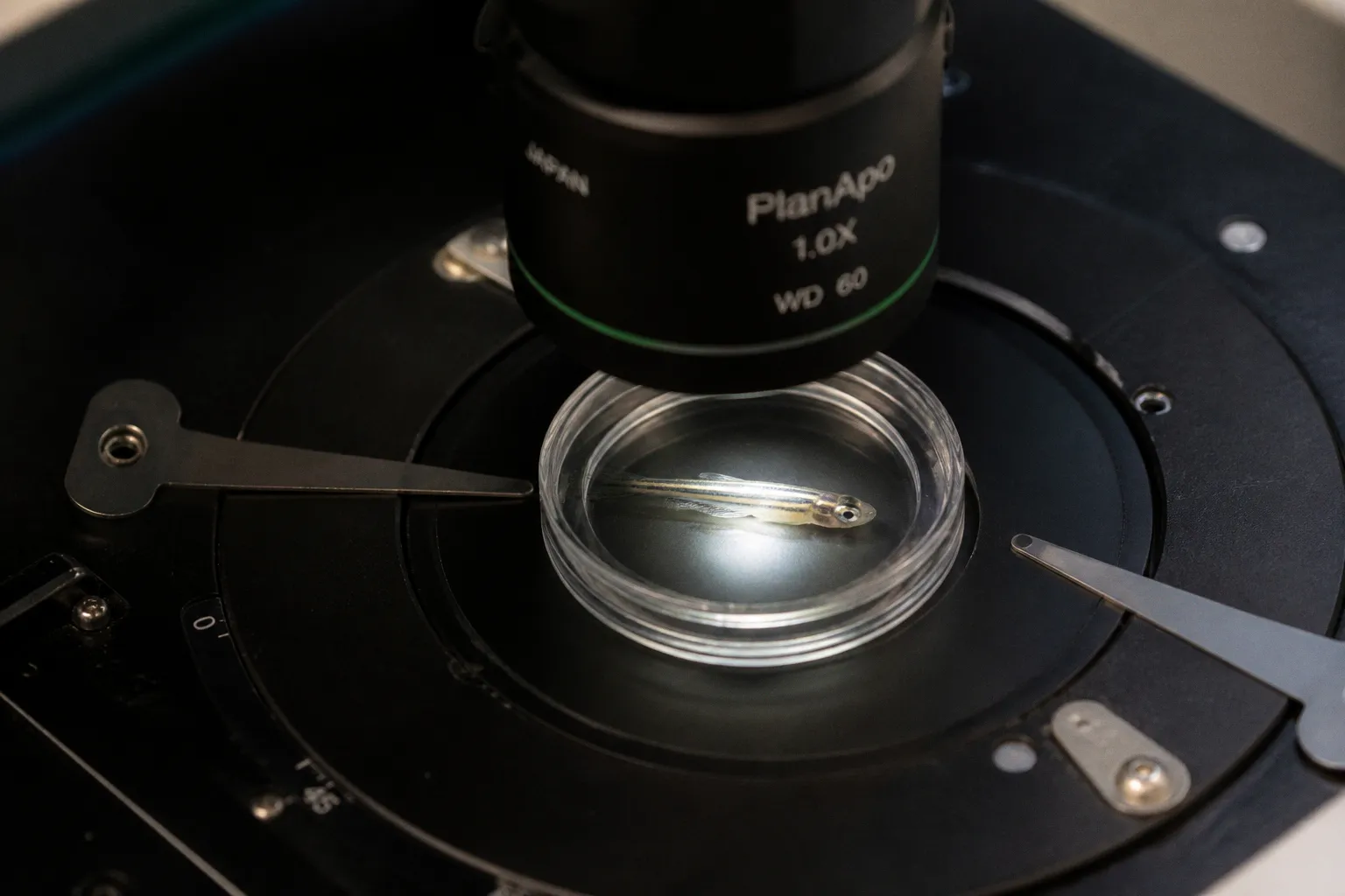
If you've ever set up a beautiful visual deprivation prep only to find your "control" larvae behaving nothing like your "experimental" larvae—and torn your hair out trying to figure out why—keep reading. According to The Transmitter, a zebrafish study published in Neuron is finally putting numbers on a hunch many of us have nursed at the bench: early visual experience doesn't just sculpt the cortex, it reshapes the retina itself, changing the shape and function of amacrine cells and altering downstream behavior. Trust me, I've watched postdocs chase that exact noise for years.
What the team actually showed
Robert Hindges's group at King's College London focused on the cells they know best—amacrine interneurons expressing the cell adhesion molecule teneurin-3, which they reported back in 2013 respond to visual stimuli aligned with the cells' physical orientation and are essential for orientation selectivity. The new work is the first to demonstrate that what an animal actually sees reshapes these interneurons themselves. Marla Feller at UC Berkeley, who wasn't involved in the study, put it bluntly: "The field in general doesn't think that activity has any effect on the retina, and it's always [acting] downstream." That framing is exactly what this paper disrupts. The retina, it turns out, is plastic in its own right, and that plasticity shows up in behavior.
Why your next prep might need a rethink
Let's talk about what this means on Monday morning. If your visual environment varies between tanks—if your "dark-reared" controls share a water system with your normally-reared animals, or your striped-stimulus rig sits next to an open bench lamp—you may not be running the experiment you think you are. The spontaneous retinal waves that start prenatally and continue until eye opening were already known to shape circuitry, but now you have evidence that patterned vision through that window rewires the cells doing the wiring. So before your next prep, let's audit three things: actual illuminance across each tank, the timing of lid opening relative to your stimulus window, and whether your control animals share any visual cues with the experimental group. Five minutes of cleanup now can save you six months of chasing phantom phenotypes later.
Don't let this become another file-drawer result
You know the feeling—beautiful paper, beautiful prep, but you can't quite get your hands around how it changes what you do tomorrow. Don't let that happen here. Pick one experiment on your bench where you suspect visual history is muddying your readout, and design a clean visual-history control this week. The tools are already on your shelf: matched LED arrays, consistent tank orientation, opaque barriers between conditions. The retina is listening. Let's make sure we're saying something worth hearing.