Chemosensory signaling before and after phosphorylation
A chemosensory receptor does not switch from “on” to “off” when its ligand disappears. The decisive control point is usually the receptor’s phosphorylation state while the ligand is still present. Kinases add phosphate groups to defined cytoplasmic sites.
Arrestins, phosphatases, adaptor proteins, and the receptor’s own intracellular loops then determine whether the signal is reduced, rerouted, or recovered.
That makes phosphorylation a timing mechanism, not a binary desensitization label. To analyze chemosensory transduction phosphorylation changes, track four variables separately: receptor occupancy, kinase access, phosphorylation-site state, and downstream output. Conflating them produces a familiar error: a drop in reporter signal is interpreted as receptor loss, even when the receptor remains at the membrane and continues to bind ligand.
In olfactory and other chemosensory GPCRs, the useful question is therefore not simply whether phosphorylation occurs. It is where it occurs, how quickly it accumulates, which molecular reader recognizes it, and what removes it.
The receptor before phosphorylation: a permissive but constrained state
Most chemosensory receptors are class A or class C G-protein-coupled receptors, although the downstream architecture differs between receptor families and cell types. Before phosphorylation, the receptor is not structurally inert. It already samples multiple conformations. Ligand binding shifts the population toward states that can engage a heterotrimeric G protein or another signaling partner.
For a canonical olfactory receptor, the first measurable sequence is:
1. An odorant binds within or near the receptor’s transmembrane ligand pocket.
2. The receptor changes the arrangement of its transmembrane helices and intracellular surfaces.
3. The receptor promotes nucleotide exchange on an olfactory G protein, commonly involving Golf in olfactory sensory neurons.
4. The G-protein pathway activates adenylyl cyclase, increasing cyclic AMP.
5. Cyclic AMP opens cyclic nucleotide-gated channels.
6. Cation influx depolarizes the neuron and recruits calcium-dependent amplification through calcium-activated chloride channels.
Phosphorylation enters this sequence on a parallel track. The same receptor activation that drives the primary signal also exposes or creates kinase-accessible states. The receptor’s C-terminal tail and intracellular loops can become substrates for G-protein-coupled receptor kinases, protein kinase A, protein kinase C, calcium/calmodulin-dependent kinases, or other context-dependent enzymes.
The exact kinase set is receptor-specific. It also depends on the cell’s baseline state, calcium load, scaffold proteins, and expression system. A receptor expressed in a heterologous cell may show a phosphorylation pattern that does not reproduce the native olfactory neuron. This is not a minor technical distinction. It changes the apparent adaptation kinetics.
A useful pre-phosphorylation model has three layers:
- Recognition: the ligand stabilizes an active receptor ensemble.
- Coupling: the active ensemble recruits the relevant G protein or effector.
- Accessibility: cytoplasmic receptor segments become available to regulatory enzymes.
The third layer is often omitted in simplified diagrams. It should not be. A ligand can produce strong G-protein activation without producing the same phosphorylation profile as another ligand. Partial agonists, inverse agonists, and biased ligands may stabilize different receptor conformations. Those conformations can differ in kinase recruitment even when their initial second-messenger output is similar.
Phosphorylation does not merely follow receptor activation. It records which active receptor state the cell has occupied.
Kinetic triggers: ligand binding starts two clocks
Chemosensory signaling adaptation is best understood as the interaction of two clocks.
The first clock measures signal initiation. It begins when the ligand-bound receptor activates its primary transducer. The second measures regulatory loading. It begins when kinases gain access to the receptor and modify its cytoplasmic sites.
These clocks can run at different speeds. If signal initiation is fast and phosphorylation is delayed, the cell produces a sharp response followed by attenuation. If phosphorylation is rapid, the receptor may enter a partially desensitized state before the downstream cascade reaches its maximum. If kinase activity depends strongly on calcium, the regulatory clock may accelerate only after the chemosensory response has already begun.
This creates several experimentally distinct states:
| Receptor state | Ligand occupancy | Phosphorylation load | Expected signaling behavior |
|---|---|---|---|
| Basal, responsive | Low or absent | Low | High capacity for new activation |
| Activated, minimally modified | High | Low | Strong initial G-protein and second-messenger output |
| Activated, phosphorylated | High | Intermediate or high | Reduced coupling, altered trafficking, or arrestin recruitment |
| Ligand-free, phosphorylated | Low | Intermediate or high | Residual desensitization after stimulus removal |
| Dephosphorylated and recycled | Low | Low | Restored responsiveness |
This table describes states, not fixed time points. A receptor can move through them quickly, remain in one state, or occupy several states at once across a cell population.
Which kinase acts first?
There is no universal order. Two broad mechanisms are common.
Homologous regulation is driven by receptor activation itself. GPCR kinases recognize features of the active receptor, often with help from membrane phospholipids or released G-protein subunits. This route tends to concentrate phosphorylation on receptors that are currently signaling.
Heterologous regulation is driven by kinases activated elsewhere in the pathway. Protein kinase A or protein kinase C can modify receptors in response to elevated cyclic AMP, calcium, diacylglycerol, or activity in neighboring pathways. This allows one signaling system to alter the gain of another.
In chemosensory neurons, calcium is a major control variable. Odorant-evoked calcium entry can activate calcium-sensitive regulatory processes, including calmodulin-dependent modulation and kinase activity. Calcium can also act downstream by changing channel behavior, phosphodiesterase activity, and chloride gradients. The observed fall in electrical response is therefore not automatically evidence of receptor phosphorylation.
To isolate receptor-level phosphorylation, separate the measurements:
- receptor surface abundance;
- ligand binding or occupancy;
- receptor phosphorylation at defined sites;
- G-protein activation;
- cyclic AMP production;
- ion-channel current;
- final membrane or behavioral output.
A single fluorescent reporter cannot resolve all seven.
Phosphorylation sites are a code, not a switch
GPCRs often contain multiple serine and threonine residues in their C-terminal tails and intracellular loops. Their modification can be sequential. One site may increase arrestin affinity. Another may change receptor conformation. A third may alter internalization or phosphatase access.
The functional result depends on the pattern. A receptor with one phosphate group is not equivalent to the same receptor with a cluster of modifications. Site order can matter because an early modification may recruit the next kinase or expose a neighboring residue. The receptor can therefore encode signaling history in its phosphorylation barcode.
For chemosensory receptors, this principle matters because odorants are rarely encountered as isolated, identical pulses in vivo. Odor concentration fluctuates. Mixtures compete. Background stimulation persists. The receptor must integrate recent exposure with current ligand occupancy. A multi-site phosphorylation system provides the necessary memory without requiring permanent receptor removal.
After phosphorylation: what actually changes
Phosphorylation can alter receptor function through several separable mechanisms. Desensitization is only one of them.
1. Reduced G-protein coupling
The most direct effect is a lower probability that the active receptor will productively engage its G protein. Phosphate groups add negative charge and alter local interaction surfaces. They can also stabilize receptor conformations that are less compatible with G-protein coupling.
This does not mean every phosphorylated receptor is inactive. Partial coupling can remain. That residual activity is often biologically useful. A cell that suppressed every receptor molecule immediately would lose sensitivity during a sustained odor plume. Graded attenuation preserves dynamic range.
2. Arrestin recruitment
Phosphorylated cytoplasmic tails and loops can recruit arrestins. Arrestin binding sterically interferes with further G-protein engagement and can promote receptor internalization. It can also create a signaling platform for kinases and other adaptors.
In olfactory systems, arrestin-mediated regulation is part of the receptor’s recovery and trafficking architecture. But the presence of arrestin does not prove that internalization has already occurred. Arrestin binding, uncoupling, clathrin recruitment, endocytosis, and recycling are distinct events. Measure them separately.
3. Changes in receptor trafficking
Phosphorylation can redirect a receptor from the plasma membrane into endocytic compartments. Internalization reduces the number of receptors available for immediate ligand detection. This is a change in receptor availability, not necessarily a permanent loss of receptor function.
The next step depends on sorting. Some internalized receptors recycle rapidly after dephosphorylation. Others remain in endosomes, where they can continue to signal through arrestin-linked pathways. Still others enter degradation routes. A bulk decrease in surface receptor signal cannot distinguish among these outcomes.
4. Altered interaction with regulatory proteins
Phosphorylation can change binding to calmodulin, scaffold proteins, phosphodiesterases, or components of the cytoskeleton. These interactions may modify receptor localization, stability, and access to downstream enzymes.
This is particularly relevant in sensory neurons, where receptor signaling is spatially organized. An olfactory receptor is not operating in a uniform cytoplasmic volume. Cilia, dendrites, and soma differ in membrane composition, ion concentration, kinase activity, and phosphatase access. A phosphorylation event in the ciliary membrane may produce a different functional effect from the same residue modified in a heterologous cell membrane.
5. Biased signal routing
A phosphorylated receptor may lose efficient G-protein coupling while retaining arrestin-dependent or scaffold-dependent signaling. Alternatively, phosphorylation can favor one G-protein pathway over another. This creates a form of signal bias that is easy to miss if the assay records only cyclic AMP.
For this reason, “desensitized” should be defined operationally. Does it mean lower cyclic AMP? Lower current? Lower calcium? Less G-protein recruitment? Reduced surface abundance? Each endpoint answers a different question.
Chemosensory signaling adaptation is not one process
Sensory adaptation occurs at multiple levels. Receptor phosphorylation is one layer in a cascade that includes second-messenger clearance, ion-channel modulation, calcium feedback, and changes in membrane excitability.
Consider an olfactory sensory neuron. An odorant activates the receptor and raises cyclic AMP. Cyclic nucleotide-gated channels open. Calcium enters. Calcium can then reduce channel sensitivity through calmodulin-dependent processes, stimulate phosphodiesterase activity, and alter other elements of the transduction machinery. The output falls even if the receptor itself remains ligand-bound and structurally active.
This produces a practical diagnostic problem. If a sustained odor reduces firing, the mechanism could be:
- receptor phosphorylation;
- arrestin-mediated uncoupling;
- cyclic AMP hydrolysis;
- cyclic nucleotide-gated channel adaptation;
- calcium-activated chloride-channel changes;
- depletion of a downstream substrate;
- altered chloride equilibrium;
- membrane excitability limits.
The solution is to perturb the system in layers. Block or reduce kinase activity, then measure receptor phosphorylation and downstream output. Separately inhibit phosphodiesterase activity. Separately manipulate calcium. Compare the time constants. A mechanism that acts upstream should change receptor-proximal measurements before it changes the final electrical response.
The same logic applies to taste GPCRs. T1R and T2R receptors detect sweet, umami, and bitter compounds through G-protein pathways, but their adaptation profile is shaped by the taste-cell environment and by downstream phospholipase C signaling, intracellular calcium, and ion-channel effects. A decrease in taste-cell output cannot be assigned to receptor phosphorylation without receptor-level evidence.
Non-olfactory olfactory receptors add another layer. These receptors appear in tissues outside the canonical olfactory epithelium and can couple to local signaling pathways that differ from Golf–adenylyl cyclase–cyclic nucleotide-gated channel logic. Their phosphorylation may regulate migration, secretion, vascular responses, or cell metabolism rather than sensory neuron firing. The receptor family name does not guarantee a shared adaptation mechanism.
Measuring phosphorylation kinetics without losing the system
The phrase “GPCR phosphorylation kinetics” hides several separate measurements. The correct assay depends on the question.
Receptor modification
Use site-specific phospho-antibodies only after validating their selectivity against phospho-null and phosphomimetic controls where feasible. Mass spectrometry can map modified residues, but sample preparation and receptor abundance impose limits. A total receptor blot cannot reveal whether the receptor population is uniformly modified or divided between unmodified and heavily modified states.
Functional coupling
BRET- or FRET-based sensors can report receptor–G-protein or receptor–arrestin proximity. Second-messenger reporters can track cyclic AMP or calcium. Electrophysiology measures the integrated output with high temporal precision.
These readouts should be aligned to the same stimulus protocol. A short odorant pulse and a prolonged bath application do not interrogate the same adaptation mechanism. Pulses isolate onset and recovery. Sustained exposure tests regulatory loading, but also changes ligand access, diffusion, and downstream resource availability.
Surface and internalized receptor pools
Label surface receptors before stimulation, then quantify loss from the membrane and return after washout. If the receptor is tagged, confirm that the tag does not alter trafficking or ligand response. Surface loss without functional recovery does not prove degradation. Functional recovery without complete surface return may indicate dephosphorylation and recycling of a small active pool.
Recovery measurements
Recovery should be measured after stimulus removal, not inferred from a lower response during the stimulus. Washout the ligand. Control the washout time. Apply a second stimulus at defined intervals. Fit the response relative to the first pulse, but report the raw traces as well.
A simple recovery experiment can distinguish at least three cases:
1. Rapid functional recovery with low surface loss: likely dephosphorylation or downstream reset.
2. Slow recovery with substantial internalization: trafficking limits receptor availability.
3. Persistent loss despite receptor return: receptor may remain modified, miscoupled, or affected by downstream adaptation.
None of these conclusions requires a single universal phosphorylation model.
If the second stimulus is weaker, do not call the receptor desensitized until surface abundance, phosphorylation state, and downstream gain have been separated.
Receptor desensitization versus signal termination
These terms describe different operations.
Signal termination stops the current response. The ligand may dissociate. The G protein may hydrolyze GTP. Cyclic AMP may be degraded. Ion channels may close. Calcium may be buffered or removed.
Desensitization lowers the receptor’s ability to respond to a subsequent or continuing stimulus. Phosphorylation is a major route into this state. It can persist after the immediate signal has ended.
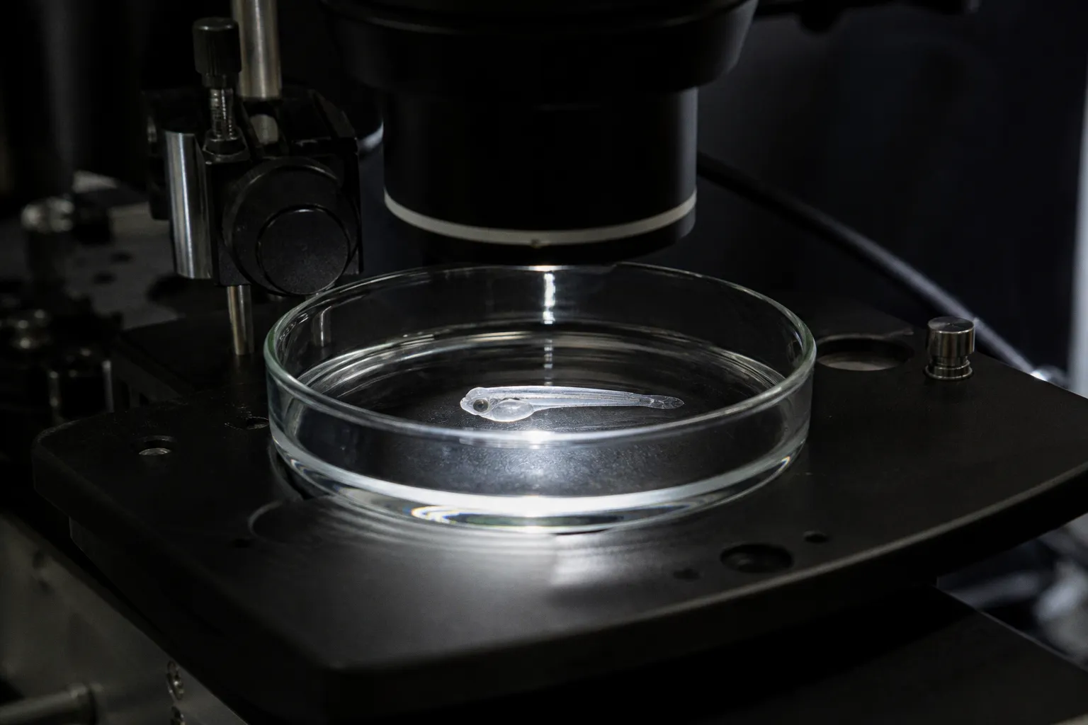
This distinction matters in behavioral sensory systems. A zebrafish may encounter an odorant concentration that changes over seconds. The receptor must terminate one event and remain ready for the next. If termination is too slow, the system integrates separate odor encounters into one prolonged response. If desensitization is too strong, the system loses contrast between background and new input.
The relevant output is not maximal receptor activity. It is temporal discrimination.
Dephosphorylation resets gain
Phosphatases remove phosphate groups and can restore receptor coupling. The reset is rarely instantaneous or complete. It depends on:
- phosphatase localization;
- receptor compartment;
- accessibility of modified residues;
- arrestin occupancy;
- ligand persistence;
- calcium and cyclic AMP levels;
- receptor recycling;
- the balance between kinase and phosphatase activity.
A receptor can therefore return to the cell surface before it returns to its original signaling competence. Surface trafficking and biochemical recovery must be measured as separate curves.
In a controlled model, receptor responsiveness can be represented as a gain variable that changes with phosphorylation load:
\[
G(t) = G_0 \times [1 - D(t)]
\]
Here, \(G_0\) is the baseline coupling gain and \(D(t)\) represents the accumulated desensitization state. A minimal balance equation is:
\[
\frac{dD}{dt} = k_{\text{phos}}A(t) - k_{\text{dephos}}D(t)
\]
\(A(t)\) is receptor activation, \(k_{\text{phos}}\) is the effective phosphorylation rate, and \(k_{\text{dephos}}\) is the recovery rate. This is not a complete molecular model. It is a diagnostic scaffold. If adaptation is stronger during high ligand occupancy, \(k_{\text{phos}}\) or the activation term may dominate. If recovery is slow after washout, \(k_{\text{dephos}}\) or receptor trafficking may be limiting.
Use this model to organize experiments, not to claim a precise molecular constant without measurements.
Why phosphorylation changes the interpretation of sensory maps
Olfactory receptor neurons are often discussed as if each receptor contributes a stable channel to the olfactory bulb. In practice, the channel gain is dynamic. Phosphorylation-dependent adaptation can change the amplitude, duration, and reliability of receptor neuron output before axons converge on glomerular targets.
That has consequences for mapping.
A receptor with high initial sensitivity but rapid phosphorylation may produce a strong onset signal and weak sustained output. Another receptor with lower initial gain but slower adaptation may dominate during prolonged exposure. The spatial map in the olfactory bulb can therefore remain anatomically stable while its temporal weighting changes.
This is not a contradiction. Wiring defines possible routes. Phosphorylation adjusts their operating range.
For mixture coding, the effect is more complex. If one odorant strongly activates a receptor and drives rapid desensitization, a second ligand acting on the same receptor may be underreported. If phosphorylation is ligand-biased, two odorants with similar initial potency can create different histories of receptor availability. Adaptation then becomes part of mixture computation.
The same principle applies to sensory processing disorders. A defect in kinase recruitment, arrestin binding, phosphatase activity, or receptor trafficking could alter perception without changing receptor expression. Expression-level assays alone will miss such defects. The system may contain the correct parts but run at the wrong gain.
Computational implications: adaptation as a local learning rule
A phosphorylation-dependent receptor can be treated as a local adaptive filter. It receives current ligand occupancy and updates its own gain based on recent activity. The receptor does not need a global representation of the odor scene. It needs only local access to activation, calcium, kinase activity, and phosphatase recovery.
This architecture provides three computational functions.
Dynamic-range compression
Strong or sustained stimuli increase phosphorylation load and reduce receptor gain. The response becomes less dominated by the most intense input. Weaker inputs remain detectable.
Temporal contrast enhancement
If adaptation tracks recent stimulation, a new change in concentration produces a larger relative response than a constant background. This is useful when the animal moves through a variable chemical environment.
History-dependent coding
The same ligand concentration can produce different outputs depending on the receptor’s prior state. That state is biochemical, not abstract. Phosphorylation, arrestin occupancy, receptor location, and downstream messenger levels collectively encode recent exposure.
A neural network model that treats each receptor as a fixed-weight input will therefore misestimate sensory output. The model should include at least one adaptive state per receptor class or per receptor population. For a minimal implementation, track:
- ligand occupancy \(L(t)\);
- receptor activation \(A(t)\);
- phosphorylation state \(P(t)\);
- effective coupling gain \(G(t)\);
- downstream messenger \(M(t)\).
The critical constraint is timescale separation. Ligand binding, G-protein activation, phosphorylation, dephosphorylation, receptor internalization, and network-level behavior do not necessarily evolve at the same rate. Compressing them into one activation variable removes the mechanism that generates adaptation.
A practical model should also allow heterogeneity. Receptor molecules within one cell may not share the same phosphorylation state. Cells expressing the same receptor can differ in kinase abundance, calcium buffering, and phosphatase activity. Population averages can conceal a fast subpopulation and a slow subpopulation, producing an apparently smooth response that no single receptor follows.
A troubleshooting sequence for phosphorylation-dependent adaptation
When a chemosensory response declines, isolate the failure point in this order:
1. Confirm ligand delivery.
Verify concentration, timing, washout, and diffusion. A weak second response may reflect incomplete stimulus control rather than receptor adaptation.
2. Measure receptor surface abundance.
Quantify the membrane pool before, during, and after stimulation. Do not infer trafficking from a functional trace.
3. Measure receptor phosphorylation directly.
Use residue-resolved methods where possible. Total receptor abundance is not a phosphorylation assay.
4. Separate G-protein coupling from downstream output.
Compare receptor–G-protein engagement with cyclic AMP, calcium, current, or firing. A downstream decline with preserved coupling points away from receptor desensitization.
5. Perturb kinase activity.
Use selective genetic or pharmacological approaches with controls for off-target changes in basal excitability and cell viability.
6. Perturb dephosphorylation and recovery.
Measure the second response after defined washout intervals. A recovery curve is more informative than one arbitrary endpoint.
7. Test arrestin and trafficking independently.
Arrestin recruitment, internalization, recycling, and degradation are not interchangeable readouts.
8. Repeat in the relevant cellular compartment.
A mechanism observed in HEK cells may not operate identically in an olfactory neuron, taste cell, or zebrafish sensory epithelium.
The strict parameter is simple: never assign adaptation to phosphorylation from a single endpoint. Require a receptor-proximal measurement, a functional coupling measurement, and a recovery measurement that agree in time.
The practical conclusion
Chemosensory receptor phosphorylation is a control layer between receptor activation and sensory output. It calibrates gain. It can isolate ongoing stimulation from new input. It can recruit arrestin, redirect trafficking, and change the receptor’s future response without removing the receptor from the membrane.
The clean analysis follows the molecular sequence: map the active receptor state, quantify phosphorylation, measure coupling, track downstream messengers, and resolve recovery after washout. Then compare the kinetics.
That sequence prevents the central interpretive mistake. A falling sensory response is not one mechanism. It is an output produced by several filters. Phosphorylation is the filter that gives the receptor memory of recent activation. To understand chemosensory signaling adaptation, measure that memory directly.