Odorant perception pathways: neural shifts before and after
A single odorant molecule contacting an olfactory sensory neuron does not enter a clean relay.
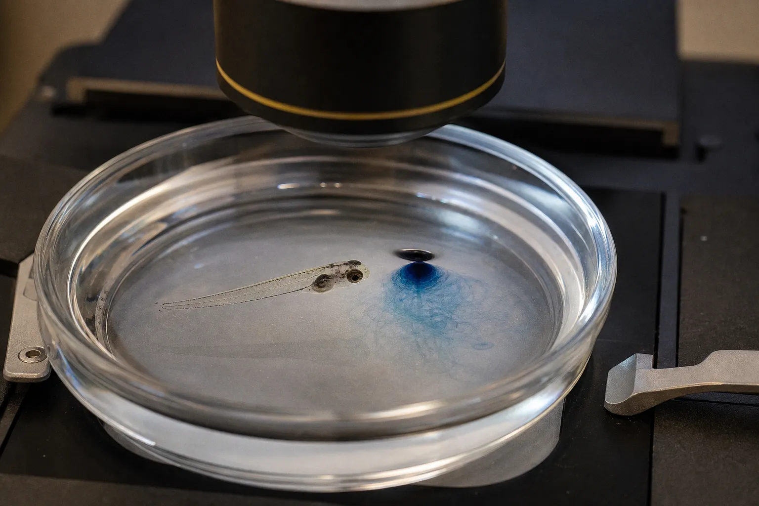
It enters a sequence of transformations that changes the signal at every stage: receptor activation becomes electrical activity, a distributed glomerular pattern becomes a more selective mitral-cell code, and the circuit may continue to represent the stimulus after the odor has disappeared.
That before-and-after difference is not a minor technical detail. It is the operating logic of the olfactory bulb. The same stimulus can look broad and chemotopic at the sensory-input stage, sparse and identity-specific at the output stage, and still leave a measurable central trace after the stimulus ends. Anyone interpreting calcium imaging in zebrafish glomeruli, mitral-cell activity in awake mice, or mixture responses therefore needs to identify where in the pathway the measurement was taken.
The phrase “odorant perception pathways” also needs to be used carefully. Olfactory sensory neurons and intranasal trigeminal neurons both detect chemical stimuli, but they do not use one universal molecular cascade. The olfactory receptor pathway is one major route; trigeminal chemosensation is a parallel system with distinct receptor classes and transduction mechanisms. Treating the two as interchangeable makes the before-and-after comparison less precise from the start.
Molecular Foundations of Chemosensory Transduction
The olfactory sensory-neuron cascade
For canonical olfactory detection, the first molecular step occurs in the cilia of an olfactory sensory neuron. An odorant receptor, a rhodopsin-like G-protein-coupled receptor, binds or is modulated by a ligand. That event initiates a signaling cascade whose central components are well characterized:
1. Receptor activation engages the olfactory-specific G protein, Golf.
2. Golf stimulates adenylyl cyclase type 3, or AC3, increasing the local concentration of cyclic AMP.
3. Cyclic AMP opens cyclic-nucleotide-gated channels, including channels containing the CNGA2 subunit, allowing cation influx.
4. The resulting calcium entry activates the calcium-dependent chloride conductance associated with TMEM16B, contributing further depolarization.
Golf, AC3, CNGA2-containing CNG channels, and TMEM16B are therefore best understood as core components of olfactory sensory-neuron transduction. They carry much of the initial signal conversion from receptor binding to membrane excitation, but the evidence does not justify describing them as an exclusive four-part chain that accounts for every peripheral mechanism. Receptor-specific behavior, channel composition, calcium handling, adaptation processes, and additional modulatory pathways can all influence the final response.
The canonical olfactory cascade is a useful experimental backbone, not a claim that every chemical-sensing neuron uses the same molecular gate.
That distinction becomes especially important when the stimulus is described broadly as “chemical” rather than specifically as an odorant presented to olfactory sensory neurons. Intranasal trigeminal chemosensation uses a different peripheral architecture. Trigeminal neurons can detect irritant, cooling, warming, burning, stinging, and pungent qualities through receptor and ion-channel mechanisms that are not reducible to the Golf–AC3–CNG–TMEM16B sequence. A stimulus can therefore produce olfactory and trigeminal signals at the same time without those signals having the same molecular origin.
What the cascade means for experimental calibration
Suppose a stimulus delivery system is working, but the signal in an olfactory-bulb glomerulus is unexpectedly weak. The right response is not to assume that the bulb has failed to process the odor. The loss may have occurred earlier: receptor engagement may be inefficient, cyclic-AMP production may be altered, calcium entry may be reduced, or the chloride-dependent amplification may contribute less than expected.
The cascade is compact enough to make this logic experimentally useful. It provides a sequence of possible bottlenecks, but not a complete inventory of all peripheral regulation. A perturbation of AC3, for example, should be interpreted as a disruption of a major olfactory transduction component, not as proof that the entire chemical-sensing periphery has a single point of failure. Likewise, reducing TMEM16B-dependent amplification can change the relationship between receptor activation and spike generation without eliminating every route by which a sensory neuron may respond.
This is also why receptor-level and glomerular-level measurements should not be treated as interchangeable. A glomerulus collects input from sensory neurons expressing related receptor identities, while the measured calcium signal reflects the combined result of receptor activation, axonal transmission, presynaptic regulation, stimulus timing, and the imaging method itself. The molecular cascade is the beginning of the pathway, not the whole explanation for what appears in the bulb.
Baseline States and the Chemotopic Organization of the Olfactory Bulb
The olfactory bulb is not simply silent until an odor arrives. Before stimulus onset, its neurons occupy a structured baseline state shaped by ongoing sensory input, internal network activity, developmental history, and recent experience. That baseline is the reference frame against which an odor-evoked change becomes visible.
In zebrafish, the olfactory bulb shows a coarse chemotopic organization. Broad classes of chemical cues tend to recruit different territories, with amino-acid-responsive inputs associated more strongly with lateral regions and bile-acid-responsive inputs associated more strongly with medial regions. The separation is useful because it gives researchers a spatial prediction before examining the finer details of individual odor responses.
| Response class | Broad olfactory-bulb territory | Interpretation |
|---|---|---|
| Amino-acid-responsive inputs | More lateral regions | Associated with a broad food-related chemical channel, while still participating in distributed representations |
| Bile-acid-responsive inputs | More medial regions | Occupy a different coarse territory rather than a completely isolated, one-odor address |
| Related or mixed stimuli | Partially overlapping territories | Reflect the distributed nature of odor coding and the convergence of receptor inputs |
This organization is already detectable early in development. Odor-evoked activity in the zebrafish olfactory bulb can be measured at approximately 2.5–3 days post-fertilization, and the broad division between amino-acid and bile-acid response territories is present at that stage. The map does not begin as an unstructured surface and then acquire all of its organization through later experience. It has an early scaffold that can subsequently be refined by activity, development, and behaviorally relevant exposure.
The word “map,” however, can be misleading. A chemotopic map is not a one-to-one lookup table in which every odor has one dedicated location. Individual odorants activate distributed sets of glomeruli, and related stimuli can recruit overlapping populations. The spatial arrangement provides a coarse organizational principle, not a complete identity label.
That matters when interpreting olfactory-bulb imaging. A broad lateral or medial response may identify the dominant chemical class of a stimulus while leaving its finer identity unresolved. Two odors can occupy a similar territory and still differ in the precise combination of glomeruli they activate, in the strength of those responses, and in their temporal evolution. Conversely, two stimuli with different chemical structures can partially converge on the same glomerular population.
The “before” frame therefore contains more than a blank baseline. It contains an organized substrate with spontaneous activity and developmental constraints. The “after” frame cannot be interpreted simply as illuminated versus non-illuminated tissue. It is a change imposed on an already active and spatially patterned network.
Temporal Reorganization: From Initial Input to Sparse Output
The first stage of odor processing is not necessarily the stage that best represents odor identity. Once sensory afferents are activated, the bulb begins to transform the incoming pattern. The transformation unfolds over time, and the time window selected for imaging or electrophysiology determines which representation is being analyzed.
A useful working sequence in zebrafish is:
1. Initial input, roughly 0–100 milliseconds. Sensory afferents generate stimulus-related activity in glomerular inputs. At this point, the response retains a strong relationship to the broad chemotopic organization: amino-acid stimuli preferentially recruit lateral regions, while bile-acid stimuli preferentially recruit medial regions.
2. Reorganization, roughly 100 milliseconds to several hundred milliseconds. Activity in initially recruited regions becomes more selective. Mitral-cell responses begin to diverge, with some cells reducing their activity, others maintaining it, and others changing their timing or response profile.
3. Transformed output. The resulting mitral-cell population pattern is sparser and more discriminative than the initial sensory-afferent representation. It is also less strictly organized by the coarse chemotopic divisions visible at the input stage.
The important point is not that the input map disappears. It is that downstream processing reads and reshapes it. In the early window, many neurons may respond in a correlated fashion because they are receiving related sensory drive. Later, inhibitory interactions and other network processes can reduce that correlation, allowing a smaller and more selective set of mitral cells to carry information about odor identity.
The olfactory bulb does not merely transmit a spatial pattern. It turns a broad input representation into a more selective, time-dependent output code.
This temporal shift is easy to miss when a recording averages an entire stimulus period into one response trace. A frame dominated by the first 100 milliseconds is answering a different question from a frame collected after the network has had several hundred milliseconds to reorganize. The first asks how sensory input arrives. The second asks how the bulb selects and distributes that input.
In practical terms, the distinction affects decoding. A classifier trained on glomerular calcium signals may learn the relatively stable spatial structure of sensory input. A classifier trained on mitral-cell activity may depend more heavily on sparse recruitment, latency, firing-rate changes, and the timing of excitation relative to inhibition. Combining these signals without preserving their temporal and anatomical origin can produce a model that appears inconsistent when the real inconsistency lies in the representations themselves.
Population size and stimulus timing also shape the measurement. Experimental work has used stimulus durations of approximately 2.4 seconds and intermediate amino-acid concentrations around 10 μM in population-imaging contexts. The adult zebrafish olfactory bulb contains an estimated 20,000 neurons, including approximately 1,500 mitral cells. These values are useful reference points for planning experiments, but they do not remove the need to report the exact preparation, imaging plane, indicator kinetics, and behavioral state. A population response is always the product of the circuit and the measurement window used to observe it.
Odor Mixture Processing and Mitral-Cell Interaction Dynamics
Mixtures expose the limits of a simple input-output model. If the olfactory bulb operated as a linear relay, the response to two odorants presented together would be close to the sum of the responses to each component. The glomerular input pattern can sometimes be predicted from the component odors within narrow limits, but that predictability does not automatically carry through to mitral-cell output.
In zebrafish, sensory afferent activity at the glomerular stage is often relatively predictable from the component patterns. At this stage, mixture responses may behave approximately additively, though “approximately” is doing important work: concentration, receptor overlap, stimulus timing, and adaptation can all affect the fit. The output stage is more complicated. Mitral-cell firing rates and response timing shift in a stimulus-dependent way, and interactions between components become stronger.
| Processing stage | What can often be predicted | Where the nonlinear change appears |
|---|---|---|
| Sensory afferents and glomerular input | Broad spatial activity from the component odors, within narrow limits | Receptor overlap, concentration, and adaptation can already modify the pattern |
| Local bulb circuitry | Which mitral cells receive convergent or competing drive | Inhibition and recurrent interactions reshape response strength and timing |
| Mitral-cell output | A selective population response rather than a simple sum | Mixture-specific temporal shifts and nonlinear firing dynamics become prominent |
The difference is conceptually important. A mixture can preserve a recognizable input structure while producing an output that is not easily reconstructed from the individual components. The bulb is not necessarily “confused” by the mixture. It is applying a transformation that changes how the components are represented together.
For decoding experiments, this creates a predictable trap. A model trained on single-odor responses may perform reasonably at the glomerular-input stage and then lose accuracy when applied to mitral-cell activity. That failure does not automatically indicate poor stimulus control or an inadequate algorithm. The model may be encountering a genuinely different coding regime in which timing and interaction terms matter.
The relevant variables include more than the number of active cells. Researchers should examine whether a mixture changes:
- the onset latency of individual mitral cells;
- the duration of their response;
- the balance between excitation and inhibition;
- the correlation structure of the active population;
- the order in which cells become active;
- and the persistence of the response after stimulus offset.
A static spatial image can conceal these changes. Two mixtures may recruit similar regions but produce different temporal sequences. Conversely, two responses that look different in their earliest glomerular patterns may converge later in mitral-cell output after local network processing.
Mixture nonlinearity is not simply noise added to the olfactory signal. It is part of the computation that gives the bulb more than a linear sum of its inputs.
This is also where the distinction between peripheral signaling and central processing becomes especially useful. Changes in sensory receptor signaling during odor exposure can alter the input pattern, but they do not by themselves explain the full mixture response. A mixture-specific shift in mitral-cell timing may arise from how convergent inputs interact with inhibition and local circuitry, even when the corresponding glomerular responses remain relatively predictable.
Central Maintenance: The Afterimage Phenomenon in Olfactory Circuits
The most striking before-and-after shift may occur after the odor is gone. In awake mice, post-odor activity has retained odor- and concentration-specific information for several breaths after stimulus offset. The circuit therefore continues to carry a representation after the physical input has ended.
This should not be described as simple peripheral persistence. Calcium-imaging and optogenetic experiments indicate that the post-odor signal is maintained primarily by central olfactory-bulb circuitry rather than by continuously active olfactory-receptor input. The bulb can preserve a stimulus-related pattern through its own network dynamics, even as the original sensory drive returns toward baseline.
During odor presentation, individual mitral cells may switch between excitation and inhibition or change the phase of their response across successive breaths. After offset, the population can continue to encode both odor identity and concentration. The signal is therefore not merely a decaying echo with no information. It behaves more like a short-lived central trace whose structure remains related to the stimulus.
That afterimage changes how “baseline” should be defined. A post-stimulus interval may be physically odor-free while remaining neurally active. If the next trial begins before the previous representation has dissipated, the measured response may include carryover from the earlier stimulus. The result can look like adaptation, increased variability, or an unexplained shift in concentration coding when it is actually a consequence of central maintenance.
Several experimental implications follow:
- The measurement window must extend beyond stimulus delivery when the question concerns persistence. Restricting analysis to the period of odor presentation misses a signal-carrying phase.
- Peripheral and central silence are not equivalent. A return of olfactory-receptor-neuron activity toward baseline does not guarantee that mitral-cell output has returned to the same state.
- Concentration information may persist alongside identity information. The afterimage is not necessarily an identity-only trace.
- Inter-trial intervals become part of the design. The timing of the next stimulus can determine whether the experiment measures a fresh response or a response shaped by the previous one.
- Species and preparation must remain explicit. Evidence for this phenomenon comes from awake mouse preparations, not from the zebrafish studies establishing early chemotopic organization and temporal reorganization.
That last distinction is more than a footnote. Zebrafish are particularly useful for optical access, developmental analysis, and mapping sensory input across the bulb. Awake mice provide a different window onto persistent activity and behaviorally relevant olfactory processing. The mechanisms may share principles, but a result demonstrated in one species should not be silently presented as established in the other.
The Trigeminal Interaction Layer
The olfactory bulb is only one part of intranasal chemical perception. The trigeminal system provides a parallel channel for sensations such as cooling, warmth, burning, stinging, pungency, and irritation. Its contribution can alter perceived intensity and behavior even when the experiment is framed as an olfactory measurement.
The two systems can be engaged by the same stimulus, but they should not be collapsed into one pathway. Olfactory sensory neurons rely on the receptor-associated cascade described above. Trigeminal chemosensation uses distinct receptors and transduction mechanisms, including pathways sensitive to irritant and thermal qualities. The fact that both channels respond to chemicals does not make their molecular gates interchangeable.
Trigeminal recruitment is also concentration-dependent and stimulus-dependent. A low-concentration odorant may produce a largely olfactory response with little detectable trigeminal contribution, while a stronger stimulus or an irritant component may activate both systems. A solvent, carrier, or delivery compound can introduce its own sensory effect. In that case, a measured response is not a pure readout of odorant perception; it is a combined olfactory–trigeminal response.
This matters at several stages:
- At the periphery, trigeminal activation can change the sensory context in which the odor is detected.
- At the bulb and connected circuits, cross-modal interactions can alter response gain and timing.
- At the behavioral level, irritation or cooling can change sniffing, withdrawal, arousal, and sampling duration.
- In data analysis, a change attributed to odor identity may actually reflect stimulus intensity or pungency.
For researchers studying sensory receptor signaling changes during odor exposure, the trigeminal channel should therefore be controlled, measured, or explicitly excluded from the claim. A clean odor-delivery protocol is not enough if the chemical stimulus itself recruits two sensory systems. Likewise, an apparent increase in olfactory-bulb activity may partly reflect altered sampling behavior caused by trigeminal sensation rather than a direct change in the olfactory code.
The safest interpretation is layered: identify which sensory neurons are being stimulated, which circuit stage is being recorded, and whether the stimulus carries a second chemical-sensing component. That approach preserves the specificity of the olfactory pathway without pretending that intranasal chemosensation has only one route.
Reading Olfactory-Bulb Changes Before and After Stimulation
A reliable comparison between pre-stimulus and post-stimulus activity begins by separating variables that are often merged into a single “odor response.” The relevant question is not only whether activity increased. It is where the activity was measured, when it occurred, and which sensory pathway could have contributed to it.
First, define the temporal frame. A signal dominated by the first 100 milliseconds is primarily informative about sensory-afferent input and initial glomerular recruitment. A signal collected 100–500 milliseconds after onset may capture the transition toward sparse, selective mitral-cell output. A post-offset signal belongs to yet another category: it may reflect central maintenance rather than continued receptor activation.
Second, keep anatomical levels separate. Glomerular calcium signals represent convergent sensory input. Mitral-cell firing reflects the result of local and network-level processing. A larger glomerular response does not necessarily predict a larger mitral-cell response, and a mixture that looks additive in the input map may become strongly nonlinear in the output pattern.
Third, treat the baseline as a measured state rather than an assumed empty interval. Spontaneous activity, recent odor history, ongoing adaptation, and post-odor afterimage can all influence the pre-stimulus comparison. If the experiment includes repeated trials, the interval between them is part of the neural protocol, not merely a scheduling detail.
Fourth, test mixtures at both stages of the pathway. A component-based model may describe glomerular input adequately while failing to predict mitral-cell timing. This is not a contradiction. It is evidence that the bulb is performing a transformation between input and output.
Finally, match the species model to the claim. Zebrafish data support detailed analysis of early chemotopic organization and temporal reorganization in the olfactory bulb. Awake-mouse data support the central afterimage phenomenon described after odor offset. Combining these findings can generate a useful comparative framework, but it should not erase the experimental boundaries between them.
The most informative before-and-after analysis is therefore not a single subtraction of two images. It is a sequence: molecular transduction in olfactory sensory neurons, spatial recruitment of glomeruli, temporal selection among mitral cells, nonlinear treatment of mixtures, and possible maintenance after the stimulus has ended. The same odorant can occupy a different representational form at each point in that sequence.
Odorant perception pathways are best understood as changing codes rather than fixed wires. Before stimulation, the bulb provides an organized and active substrate. During the first moments of exposure, sensory input establishes a distributed chemotopic pattern. Over the next several hundred milliseconds, local circuitry reshapes that pattern into a sparser output. After offset, central dynamics may preserve an odor- and concentration-related trace. And when trigeminal input is present, a parallel chemical-sensing system can modify the entire trajectory.
That is the practical meaning of the neural shift before and after activation: the experiment does not observe one response moving through a tube. It observes successive versions of the stimulus, each produced by a different layer of the circuit.