Trigeminal receptor signaling: changes after capsaicin
A single bolus of capsaicin injected into deep craniofacial tissue drops the mechanical activation threshold of trigeminal nociceptive afferents almost immediately. That is the entry point. Follow the signal longer—minutes, hours, days—and the system inverts.
The same neurons that fired intensely under the initial stimulus enter a refractory state in which they barely register nociceptive input. The mechanism behind that inversion is not mysterious. It is quantifiable, mapped at the molecular level, and reproducible across experimental preparations.
The important distinction is temporal. Trigeminal receptor signaling before and after capsaicin is not one stable state with one predictable output. It is a sequence: activation, sensitization, calcium-dependent suppression, receptor trafficking, neuropeptide downregulation, and gradual recovery. The receptor does not simply turn on and then turn off. The entire sensory neuron changes its operating state.
TRPV1 Activation and the Initial Nociceptive Cascade
Capsaicin binds to TRPV1, a nonselective cation channel expressed on trigeminal nociceptive afferents. These are sensory neurons, not motor neurons and not proprioceptors. TRPV1 expression is selective, although the receptor is also found on nociceptive fibers in other parts of the peripheral nervous system. Once capsaicin occupies its binding site, it forces a conformational change that opens the channel pore.
The immediate result is an influx of calcium and sodium. The membrane depolarizes, voltage-gated channels amplify the disturbance, and action potentials propagate toward the trigeminal ganglion and onward to the trigeminal nucleus caudalis in the brainstem. The organism perceives burning pain, heat, and irritation. At the peripheral terminal, the same activation can drive vasodilation and local inflammatory signaling.
This is not a diffuse process. In cultured trigeminal ganglion neurons, capsaicin activates two distinct inward currents: a slow current and a rapid current. The rapid current requires extracellular calcium. It is insensitive to resiniferatoxin, a related TRPV1 agonist, which indicates that the two responses partially diverge at the receptor or channel-regulation level. The study identifying this dual-current architecture dates to 1994 and used rat trigeminal preparations, but the distinction remains useful because it shows that "TRPV1 activation" is not a single uniform electrical event.
Before desensitization enters the picture, the initial cascade can be described in five linked stages:
1. Ligand binding. Capsaicin occupies the vanilloid binding pocket on TRPV1.
2. Channel opening. A conformational shift permits calcium and sodium influx.
3. Depolarization. The membrane potential changes, allowing voltage-gated channels to propagate the signal.
4. Neurotransmitter release. Substance P, CGRP, and somatostatin are released from peripheral and central terminals.
5. Sensitization. Peripheral injection of 1% capsaicin into deep craniofacial tissues sensitizes nociceptive afferents and lowers their mechanical activation threshold.
The fifth stage is easy to miss because capsaicin is often discussed mainly as a desensitizing agent. Its first effect is the opposite. The neuron becomes more responsive. Mechanical input that would previously have remained below threshold can now produce firing, and the same stimulus can feel more intense when delivered repeatedly over a short interval.
That initial sensitization is the starting condition for everything that follows. Calcium-dependent desensitization, receptor internalization, and neuropeptide depletion are not independent processes added from outside. They are compensatory responses to the intensity and duration of the initial TRPV1 signal.
Capsaicin does not block trigeminal signaling at the outset. It drives the system hard enough that the system begins to suppress its own output.
The distinction matters experimentally. A response measured seconds after capsaicin exposure may represent channel activation and peripheral sensitization. A response measured later may reflect dephosphorylation, depletion of regulatory lipids, receptor trafficking, or changes in gene expression. The same preparation can therefore produce opposite results without any contradiction in the underlying biology.
Molecular Mechanisms of Calcium-Dependent Desensitization
Desensitization is not a single event. It is a cascade of molecular adjustments triggered by the same calcium influx that initiates nociception. TRPV1 activates mechanisms that progressively reduce its own activity.
From calcium entry to reduced channel activity
The first stage is calcium sensing. Elevated cytosolic calcium binds calmodulin, a calcium-sensitive regulatory protein present in neurons. Calmodulin then participates in the activation of calcineurin, a calcium-dependent phosphatase.
Calcineurin dephosphorylates TRPV1 at regulatory serine and threonine residues. Phosphorylation generally increases the channel's readiness to open or helps maintain its sensitized state. Dephosphorylation has the opposite effect: the probability of channel opening falls, and subsequent capsaicin stimulation produces a weaker current.
Calcium also activates phospholipase C delta, or PLCδ. This enzyme hydrolyzes phosphatidylinositol 4,5-bisphosphate, commonly referred to as PIP2, in the plasma membrane. PIP2 is not simply structural membrane material. It acts as a regulatory lipid that helps sustain TRPV1 activity. When PIP2 is depleted, channel conductance declines further.
The sequence is therefore not a linear switch but a set of overlapping brakes:
- Calcium enters through the activated channel.
- Calmodulin detects the rise in intracellular calcium.
- Calcineurin removes phosphate groups from TRPV1.
- PLCδ reduces the local supply of PIP2.
- The channel becomes less likely to remain active.
- Further capsaicin stimulation produces a smaller response.
The critical parameter is extracellular calcium. Remove it from the bathing medium and the expected desensitization does not develop normally. This was demonstrated directly in trigeminal ganglion preparations. The initial capsaicin response can still be observed under some experimental conditions, but the subsequent refractory state is substantially altered when calcium entry is prevented. Calcium is therefore not just a charge carrier that initiates the signal. It is also the trigger for the feedback mechanisms that suppress it.
| Molecular event | Trigger | Downstream effect |
|---|---|---|
| Calmodulin activation | Rise in cytosolic Ca²⁺ | Recruitment of calcium-sensitive regulatory pathways |
| TRPV1 dephosphorylation | Calcineurin activation | Lower channel open probability |
| PIP2 hydrolysis | Ca²⁺-activated PLCδ | Reduced support for sustained TRPV1 activity |
| Receptor endocytosis | Prolonged or repeated stimulation | Removal of TRPV1 from the cell surface |
| Neuropeptide transcriptional shift | Sustained capsaicin exposure | Reduced capacity for later neuropeptide production |
The temporal resolution matters. Calmodulin activation, calcineurin signaling, and PIP2 depletion operate on the scale of minutes. They alter the behavior of receptors that are still present in the membrane. Endocytosis develops more slowly and produces a more durable change: the receptor is no longer available at the cell surface to detect the next stimulus.
This is the molecular basis of trigeminal nerve desensitization. It is not equivalent to neuronal death, and it is not the same as cutting off conduction. The axon remains present. The neuron can still respond through other channels and transmitters. What changes is the gain of a particular nociceptive pathway and the ability of the cell to sustain that gain after strong stimulation.
Receptor Internalization and Neuropeptide Expression Shifts
Prolonged capsaicin stimulation does not merely silence TRPV1 at the membrane level. It can remove the receptor from the membrane altogether.
Continuous activation induces endocytosis: TRPV1 is pulled from the cell surface into intracellular vesicles and subsequently trafficked through sorting and degradation pathways, including lysosomal processing. The neuron temporarily loses part of its primary detection apparatus for noxious thermal and chemical stimuli. Recovery then requires the synthesis and delivery of new TRPV1 protein. Re-phosphorylation of the receptors that remain at the membrane is not enough.
That is a structural intervention rather than a simple change in channel tuning. The neuron is physically reorganized at the level of its receptor inventory. The result is slower to appear than acute calcium-dependent desensitization, but it can last longer because the cell must replace what has been internalized.
Capsaicin also changes the neuron's signaling output at the transcriptional level. In cultured trigeminal ganglion neurons, exposure reduces mRNA expression of several neuropeptides:
- Substance P: a 2.07-fold decrease
- CGRP, or calcitonin gene-related peptide: a 3.09-fold decrease
- Somatostatin: a 1.60-fold decrease
The CGRP result is especially important. A reported 3.09-fold decrease in CGRP mRNA indicates a strong reduction in transcript abundance under the experimental conditions; it should not be converted into an unsupported percentage reduction in the neuron's inflammatory signaling capacity. mRNA abundance is one layer of regulation. It does not translate automatically into an identical change in peptide release, receptor signaling, or tissue-level inflammation. The finding is still substantial, but its meaning must remain attached to what was actually measured.
CGRP is a major mediator of neurogenic inflammation and an established therapeutic target in migraine pharmacology. A reduction in its transcript can limit the cell's later capacity to replenish peptide stores, particularly when exposure is sustained or repeated. Substance P and somatostatin shift as well, but not to the same degree in this preparation. The response is therefore not a generic shutdown of all neuronal gene expression. It is a selective remodeling of molecules that contribute to nociceptive and inflammatory communication.
Peptide depletion is not the same as loss of nociception
The historical data on substance P makes this point clearly. Neonatal capsaicin pretreatment in rats depletes substance P levels in the trigeminal nucleus caudalis by 55.6%. One-month postnatal treatment achieves 57.9% depletion. Both protocols reduce thermal nociceptive responses. But adult pretreatment with the same molecule does not reduce thermal responses to the same degree, despite comparable substance P depletion.
The implication is not that substance P is irrelevant. It is that substance P is not the sole transmitter mediating thermal nociception in this circuit. Other transmitters and pathways can compensate, and the capacity for compensation changes with developmental state. A molecular marker can therefore show a pronounced shift while the behavioral phenotype remains partial.
This is one reason the phrase "capsaicin sensory receptor changes" needs to be handled carefully. There are at least three different measurements that may be described under that heading:
1. Channel availability: how much TRPV1 remains at the membrane.
2. Channel responsiveness: how strongly the remaining receptor responds to capsaicin or heat.
3. Neuronal output: how much neuropeptide and electrical activity the neuron can produce after exposure.
These measurements move together only during part of the response. A neuron may have desensitized membrane receptors while retaining the ability to conduct signals through other channels. It may show reduced neuropeptide mRNA without an immediate equivalent reduction in electrically evoked transmission. The before-and-after comparison is meaningful only if the measurement and time point are specified.
The neuron does not simply go quiet. It removes receptor machinery from the membrane and changes the molecular supply lines behind its pain signal.
Regional Variations in Trigeminal Dermatome Sensitivity
The trigeminal nerve divides into three primary branches: V1, the ophthalmic division; V2, the maxillary division; and V3, the mandibular division. Each innervates a distinct facial dermatome, and capsaicin does not produce uniform responses across them.
Topical capsaicin application changes somatosensory sensitivity in a branch-dependent manner. Applied to the nasal mucosa, which receives mixed trigeminal innervation from both V1 (primarily the anterior ethmoidal nerve supplying the anterior nasal cavity) and V2 (the nasopalatine and lateral posterior nasal branches supplying the posterior nasal cavity), it triggers robust local and generalized parasympathetic activation, including lacrimation and increased secretory output. The response is not limited to conscious nociception. It recruits autonomic pathways through a trigeminal relay.
That parasympathetic recruitment does not respect the anatomical branch boundaries that divide the rest of the dermatome. A mucosal stimulus can drive lacrimation, nasal secretion, and conjunctival output together, because the efferent limb runs through parasympathetic ganglia that are activated regardless of which afferent branch carried the signal in. The mixed afferent supply at the nasal mucosa means that branch assignment should not be treated as the determinant of the reflex outcome.
Vascular responses also show regional asymmetry. Topical capsaicin increases dermal blood flow in trigeminal dermatomes at a significantly higher rate than in extracephalic control sites such as the forearm. The difference is consistent with the dense involvement of trigeminal perivascular afferents and with the role of CGRP in capsaicin-mediated vasodilation.
The anomaly appears in migraine. After identical capsaicin exposure, patients with migraine show a smaller increase in dermal blood flow in the V1 dermatome than healthy controls. The mechanism is not fully resolved. It could reflect altered receptor density, pre-existing changes in CGRP availability, vascular wall adaptations, or a combination of these factors. The available data do not establish one explanation conclusively.
What the result does establish is more useful than a premature mechanism: trigeminal sensitivity to capsaicin is not a single variable. It depends on the branch being tested, the tissue being stimulated, the history of prior activation, and the neurological state of the subject. A response in V1 skin cannot automatically be treated as a proxy for the response of V2-innervated facial regions, and neither can be treated as a proxy for the mixed V1/V2 territory of the nasal mucosa. Regional attribution has to match the actual innervation pattern of the tissue under study.
Nasal chemosensation and trigeminal relay
The nasal mucosa deserves separate attention because it exposes the difference between nociceptive signaling and reflex chemosensation. Here, capsaicin acts on sensory nerve endings that overlap functionally with the trigeminal nociceptive system but are not identical to every class of trigeminal afferent.
The resulting parasympathetic outflow—lacrimation, secretion, and related glandular responses—can occur without being experienced as proportional conscious pain. This is a chemosensory transduction pathway operating through trigeminal circuitry. It is a crossover point where irritation, autonomic reflexes, and sensory detection converge on the same broad anatomical system without becoming the same signal.
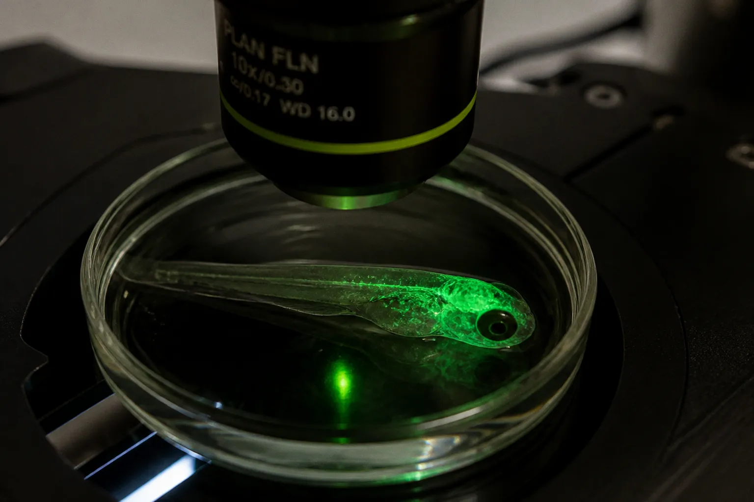
That distinction also matters for zebrafish and other experimental models of sensory neurobiology. A receptor response observed in an isolated sensory neuron may reveal the channel mechanism, while an intact-organism response includes ganglionic integration, central projections, autonomic reflexes, and local vascular effects. The model is not a miniature version of the human face; it is a way to separate levels of the pathway and ask which part changes first.
Temporal Dynamics: From Sensitization to Refractory States
Timing controls the outcome. The same stimulus produces opposite effects depending on when the response is measured.
Apply capsaicin to the tongue at a one-minute interstimulus interval and the irritant sensation becomes stronger with each application. The system is sensitized, not suppressed. The interval is short enough for the ongoing excitability of the afferent and its local inflammatory output to reinforce the next response.
Extend the interval and the trajectory reverses. Calcium-dependent dephosphorylation, regulatory-lipid depletion, and receptor endocytosis accumulate across the gap between stimuli. Each successive application lands on a neuron that has already begun to dismantle its detection apparatus. The net psychophysical rating drops, and the channel current recorded from isolated neurons under matched stimulation falls in parallel.
The same applies at the dermatome level. Repeated topical application across days does not just lower the response to capsaicin itself; it lowers the baseline sensitivity to mechanical and thermal stimuli in the surrounding skin. That prolonged suppression is the rationale behind capsaicin-based topical therapeutics, and it is also the reason the molecule has to be treated as a state-changing agent rather than a simple blocker.
Two practical corollaries follow for anyone designing or interpreting experiments:
- Short-interval protocols sample the sensitization phase, not desensitization. They reflect the early part of the cascade, where calcium influx drives increased excitability and neuropeptide release.
- Long-interval and repeated-application protocols sample the refractory phase. They reflect the cumulative effect of dephosphorylation, lipid depletion, receptor internalization, and transcriptional shifts.
The reversal is reproducible, but its boundary depends on concentration, route, tissue, species, and developmental stage. A 1% deep-tissue injection in an adult rat and a low-dose topical application on human forearm skin are not the same preparation, even when the readouts look superficially comparable.
That is the working definition of trigeminal receptor signaling before and after capsaicin: a reversible, time-locked, region-specific inversion of sensory gain. It is not a switch that turns the nerve off. It is a graded reconfiguration of the detection machinery that runs from channel gating through gene expression, with the same neuron holding all of those layers at once.