Larval zebrafish motor recovery after spinal cord injury
A mechanical lesion in a three-day-old zebrafish larva closes within roughly forty-eight hours. Within several days after that seal, the same larva may respond to touch, execute coordinated escape movements, and resume free swimming.
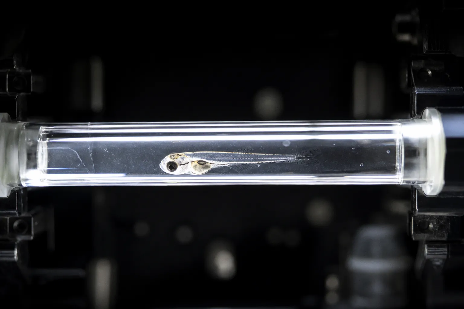
That speed is one reason the larval zebrafish has become such a useful functional recovery zebrafish model. It is also the source of a recurring interpretive error: movement returning is not automatically the same thing as the original spinal circuitry being restored.
The experimental question is therefore not simply whether a larva swims again. It is which component of sensorimotor function has returned, on what timescale, and how that behavioral change relates to the anatomical events visible at the lesion. That distinction shapes every protocol choice, from injury staging and imaging intervals to the selection of a locomotor assay.
Temporal Dynamics of Functional Restoration: From Sensory Return to Free Swimming
Recovery after larval spinal cord transection follows a staged sequence, but the sequence is not identical across assays or developmental stages. The timeline compresses what takes months in mammals into roughly a week, while the readout can change the apparent speed and completeness of recovery by several days.
In a 5 dpf complete-transection model, sensory responsiveness measured through touch-evoked reactions returns by 2 days post-injury (dpi). This is the earliest reported functional signal in the sequence. C-bend escape movements, which require coordinated bilateral motor output, become detectable at 3 dpi. Free swimming resumes by 5 dpi. In a separate 5 dpf analysis using total distance moved and turn angle, full locomotor recovery was reported by 6 dpi relative to age-matched uninjured controls.
These are not interchangeable endpoints. A touch-evoked response can be generated by a relatively fast sensory-to-motor pathway without demonstrating that the larva can sustain normal exploratory swimming. Conversely, total distance moved may look close to control values while turning remains abnormal or the animal relies on a less precise motor strategy. The label "recovered" only becomes useful when the assay behind it is stated clearly.
The 10 dpf transection paradigm gives a more restrained picture. In this model, the functional readout was trunk curvature during a dark-flash visual motor response. Immediately after injury, the response rate fell to 3.0 ± 1.5%, compared with 54.3 ± 4.6% in controls. Partial recovery followed, reaching 28.6 ± 5.6% at 2 dpi. By 4 dpi, the rate had plateaued at 24.5 ± 6.0%, still below half of the control value of 59.2 ± 5.4%. Video acquisition ran at 1,000 frames/s for one-second windows during each dark-flash trial.
The contrast is informative, but it should not be reduced to a simple statement that young larvae regenerate and older larvae fail. The 5 dpf and 10 dpf experiments differ in age, lesion context, assay, stimulus pathway, and observation window. The result is best read as an age-dependent difference in the recovery profile measured by those particular paradigms.
Recovery timing is partly a biology readout and partly an assay decision. A caudal tail-touch response can return days before a visually evoked motor response approaches its control level.
For a screen aimed at spinal repair, the behavioral endpoint should be chosen before the injury age is fixed. Otherwise, the protocol may produce a clean and reproducible curve that answers a different question from the one the experiment was designed to address.
What the early signals do—and do not—show
The first returning response is not necessarily the most informative one. Sensory responsiveness establishes that some pathway from stimulus detection to motor output is operating again. It does not establish normal descending control, accurate left-right coordination, or sustained swimming. Likewise, free swimming establishes a broader level of motor function but can conceal changes in bout structure, turning, speed, and body curvature.
A useful recovery series therefore separates at least three levels:
- Stimulus-evoked responsiveness, which is sensitive to the return of a rapid sensory-motor pathway.
- Coordinated escape, which places greater demands on bilateral motor output and timing.
- Voluntary or visually evoked locomotion, which engages broader circuit integration and may expose deficits hidden by a reflex assay.
The same larva can appear recovered at one level and impaired at another. That is not a contradiction. It is the expected consequence of measuring different parts of the locomotor system.
Anatomical Reorganization: Axonal Regrowth and Lesion Bridging Mechanisms
The anatomical basis of larval zebrafish spinal cord regeneration is often described too confidently. Wound closure, axonal extension, and bridging are observable events. Synaptic specificity and full circuit reconnection are stronger claims and require different evidence.
In the 10 dpf transection model with GFP-labeled axons, isolated axons were observed entering the lesion site by 24 hours post-injury. By 48 hours, substantial longitudinal axon bundles bridged the transection. These observations provide direct evidence of axonal entry and lesion bridging in the imaging preparation. They do not, by themselves, demonstrate that the axons have re-established their original synaptic partners or that the reconstructed pathway produces normal circuit output.
The distinction is central to zebrafish axon regrowth imaging. A bundle that spans the lesion may contain axons with different destinations, growth states, and degrees of organization. Some may extend through a permissive route without recreating the original connectivity. Intravital imaging can show where labeled axons go and when they cross the lesion; it cannot alone establish synaptic reconnection unless paired with anatomical tracing, synaptic markers, physiological measurements, or another circuit-level assay.
The mechanical lesion in the 3 dpf protocol closed within roughly 48 hours as well. Wound closure and axonal bridging therefore occur on overlapping, but not identical, timescales. A sealed wound is not evidence that the tissue has regained its pre-injury architecture. Similarly, axonal bridging should be treated as a structural milestone rather than the final definition of repair.
The 2016 work on motor neuron regeneration established that larval spinal motor neurons can be regenerated after both mechanical lesion and genetic ablation. When the immune response was suppressed with dexamethasone, motor neuron regeneration decreased. That result gives the field an important pharmacological comparison: immune activity is not merely an aftermath of injury but can influence the regenerative capacity of the motor circuit. It still does not mean that every immune response is beneficial, or that the regenerated neurons necessarily restore the original connectivity.
Building an anatomical time course
A useful imaging series begins with a fixed injury age. The 3 dpf, 5 dpf, 7 dpf, and 10 dpf paradigms are not interchangeable: they differ in developmental state, cellular composition, inflammatory kinetics, and the apparent dependence of axons on glial structures.
For intravital confocal work, 2% low-melting-point agarose is a standard immobilization medium. At the 10 dpf stage, the transection and imaging procedures described in the protocol use 0.015% buffered tricaine. The exact timing of image acquisition matters as much as the imaging label. Intervals at 6, 12, 24, and 48 hpi can capture the transition from early axonal entry to more substantial bridging, provided the same anatomical landmarks and analysis rules are maintained.
The most informative design pairs those structural time points with behavioral measurements. An image showing axons crossing the lesion should not be presented as a functional endpoint unless the corresponding motor data support that interpretation. The strongest result is often a partial alignment: structural bridging begins before a complex behavior returns, or gross movement returns while anatomical organization remains visibly immature.
That apparent mismatch is not experimental failure. It is the point at which neural circuit reorganization post-injury becomes experimentally visible.
The Role of Innate Immunity: Macrophage Dynamics in Spinal Repair
The immune system is not a background variable in larval spinal repair. It helps determine whether the lesion environment becomes permissive for regrowth, and different innate immune populations cannot be treated as interchangeable.
Peripheral macrophages are implicated in complete functional recovery after larval spinal cord injury. Macrophage-deficient models show prolonged inflammation and impaired repair. The relevant mechanism is unlikely to be limited to debris clearance. Macrophage activity can alter the cellular environment around the lesion, influence the persistence of inflammatory signals, and help create conditions in which axons can extend across damaged tissue.
Neutrophils, by contrast, are dispensable for spinal cord regeneration in this context. That asymmetry is experimentally important. A manipulation that changes macrophage recruitment or function is not equivalent to one that changes neutrophil abundance. If the experiment uses a pan-leukocyte marker, it may show that immune cells are present while failing to identify the population associated with the regenerative phenotype.
The dexamethasone result reinforces the point from another direction. Broad immunosuppression reduces motor neuron regeneration. This does not establish that inflammation is uniformly protective. It indicates that suppressing immune activity without preserving the relevant regenerative component can impair the outcome. The useful interpretation is more specific: the timing and identity of the immune response matter.
In larval spinal repair, "the immune response" is too broad a category to be a mechanism. Macrophage dynamics and neutrophil dynamics carry different experimental meanings.
For a compound that modulates immune function after injury, macrophage recruitment should be tracked separately rather than inferred from a general leukocyte signal. Recruitment alone is also not enough. Cell number, residence time, localization relative to the lesion, and the timing of depletion or suppression can all alter the phenotype. A macrophage population present at one time point may not perform the same role as one present later in the repair sequence.
This is where functional and anatomical measurements need to meet. If a treatment increases the number of macrophages at the lesion but does not improve axonal bridging or locomotor output, the result is not evidence of successful repair. It may indicate altered recruitment without productive tissue remodeling. Conversely, a behavioral improvement in the absence of a measurable change in bridging could reflect compensation or a pathway not captured by the chosen marker.
Age-Dependent Plasticity: Comparing Injury Responses Across Larval Stages
The comparison across 3, 5, and 7 dpf injury ages in the 2025 systematic study clarified a point that earlier observations had suggested: larval age at injury is not a minor protocol parameter. It changes the regenerative context in which every subsequent measurement is made.
Younger larvae at 3 dpf regenerated axons more independently of a glial bridge. In practical terms, the regenerating axons were less dependent on scaffold cells to guide them across the lesion. Older larvae at 7 dpf showed a different pattern, including:
- More sustained cell death at and around the lesion site
- Delayed inflammatory responses
- Different glial bridging patterns
- Greater dependence on glial scaffolds for axonal crossing
The comparison is useful because it separates two questions that are often collapsed into one. If the aim is to study axon-intrinsic regenerative capacity, a younger stage may reduce the contribution of glial scaffolding. If the aim is to understand glial-dependent axon guidance, that same stage may be a poor choice because the mechanism under study is less prominent.
Age also changes what counts as a convincing behavioral endpoint. The 3 dpf mechanical-lesion protocol produced swimming recovery within approximately two days when assessed by caudal tail-touch escape. The 5 dpf transection protocol showed free swimming by 5 dpi and C-bend escape by 3 dpi, while a separate analysis of total distance moved and turn angle reported full locomotor recovery by 6 dpi. The 10 dpf dark-flash assay did not reach control-level performance within the observation window.
These results should be compared as recovery trajectories, not arranged into a universal ranking of "good" and "bad" ages. A larva injured at 3 dpf is still undergoing rapid development. Its motor system, body size, tissue geometry, and baseline behavior are changing alongside the repair process. The 10 dpf animal presents a different substrate, with a more established circuit and a different lesion response. A slower or incomplete behavioral recovery may therefore reflect both reduced regenerative plasticity and a more demanding functional test.
Here is the practical comparison across the published paradigms:
| Parameter | 3 dpf | 5 dpf | 10 dpf |
|---|---|---|---|
| Lesion type | Mechanical | Complete transection | Transection |
| Lesion closure | Approximately 48 hours | Not specified in the cited comparison | Not specified in the cited comparison |
| First axonal entry | Not specified | Not specified | 24 hpi |
| Axonal bridging | Not specified | Not specified | 48 hpi |
| Sensory recovery | Approximately 2 days (tail-touch response) | 2 dpi (touch-evoked) | Not specified |
| Motor recovery | Approximately 2 days in a tail-touch assay | Free swimming at 5 dpi; C-bend escape at 3 dpi; full locomotor recovery at 6 dpi in turn angle and distance assays | Partial recovery at 4 dpi in the dark-flash response |
| Key assay | Tail-touch escape | Free swimming, turn angle, C-bend escape | Dark-flash visual motor response |
| Glial dependence | Lower in the 3–7 dpf age comparison | Not specified | Not specified in the cited comparison |
The through-line is not that young tissue simply repairs itself and older tissue does not. Younger tissue follows a faster, less glial-dependent program in the cited comparisons. Older tissue shows more sustained cell death, altered immune timing, and a stronger dependence on glial scaffolds, with incomplete restoration in the tested behavioral window.
That distinction matters when interpreting a candidate regenerative treatment. A compound that improves axonal bridging at 7 dpf may be acting on a mechanism that is barely engaged at 3 dpf. Conversely, an intervention that appears ineffective in an older animal may still influence early structural events without being sufficient to restore a demanding visual motor response.
Quantitative Behavioral Assays: Measuring Locomotor Recovery in the Lab
Perfectly registered anatomy can still produce a misleading recovery curve if the behavioral assay is poorly matched to the circuit being studied. The central issue is not whether an assay is quantitative. It is whether its measured variable is close enough to the biological question to support the conclusion being drawn.
A validated open-source MATLAB method quantifies trunk curvature during propulsive and turning movements in larval zebrafish. The method is designed for simultaneous analysis of multiple larvae housed in separate chambers. That throughput is relevant to large injury experiments: the 5 dpf micropipette transection protocol reports a throughput of 300 larvae per hour, with 98.75% ± 0.72% survival to 7 dpi across more than 3,600 transected larvae.
That survival result indicates low attrition under the reported conditions. It does not prove that the mechanical procedure introduces no confounding or that every surviving larva received an identical lesion. Throughput numbers belong to the protocol, not to the lesion: a fast and reproducible transection is a tool, not a guarantee of uniform tissue damage.
The choice of variable still drives interpretation. Total distance moved captures how far the larva travels but says little about how the swim is produced. Turn angle captures the geometry of directional changes but is silent on bout structure. Trunk curvature captures the bend pattern that produces propulsion and turning and is therefore closer to the underlying motor output. A free-swimming recovery curve that integrates only distance can flatter a larva whose swim pattern remains abnormal.
For an experiment designed to measure locomotor function restoration in zebrafish, the assay should be specified before the lesion is made. The same injury paradigm can produce three different recovery curves depending on whether the readout is touch-evoked response, C-bend escape, total distance, or trunk curvature during a visual motor response. None of these is wrong in isolation. The error is in treating them as interchangeable.
Putting the assays together in a single study
A coherent recovery study pairs at least one structural measure with at least one behavioral measure across matched time points. The combination that has worked in the published literature is:
- A 3 dpf or 5 dpf injury with a fixed transection or mechanical lesion protocol
- Repeated anatomical imaging at 6, 12, 24, and 48 hpi to capture axonal entry and bridging
- A touch-evoked response test at 1–2 dpi to capture the earliest functional signal
- A C-bend escape assay at 3 dpi to confirm coordinated motor output
- Free swimming and trunk curvature measurements from 5 dpi onward to capture the consolidation of recovery
When an intervention is added, the same set of measurements should be applied to treated and control animals. A treatment that improves axonal bridging without accelerating any behavioral endpoint is a structural result, not a functional one. A treatment that improves behavior without changing bridging is a functional result that needs an explanation through another circuit-level measurement. The interpretation depends on which of these pairs is reported.
Matching the Protocol to the Question
The choice of injury age, lesion type, and assay defines the experiment more than any single reagent or transgenic line. A short summary of the practical tradeoffs:
- 3 dpf mechanical lesion: fast, reproducible, and useful for axon-intrinsic regeneration questions. Limited strength of the behavioral readouts because the larva is still developing rapidly.
- 5 dpf complete transection: better defined behavioral assays, including free swimming and C-bend escape, and a more mature motor system. Useful for screens where functional endpoints are the primary readout.
- 7 dpf transection: more sustained cell death and clearer glial dependence, which is useful for studying scaffold-guided regeneration. Behavioral recovery is slower and may not reach control levels within the standard window.
- 10 dpf transection: a more demanding functional test (dark-flash visual motor response) and an established baseline for incomplete recovery. Useful for studying why behavioral restoration can fail even when structural bridging is present.
The label "zebrafish model" covers four different experimental contexts. Treating them as a single system is the most common source of contradictory conclusions in the literature.
The strongest use of this model is not to ask whether larval zebrafish "regenerate." It is to ask which component of recovery, in which lesion context, and through which assay, has been moved by the experimental intervention. With that framing, the speed of the system becomes a tool rather than a distraction, and the zebrafish spinal cord regeneration mechanisms under study stay attached to the specific behavior they are meant to explain.