Non-olfactory olfactory receptors: 5 ways they shape sensory input
The nose was never the whole story. For decades, olfactory receptors were treated as a closed system: molecular sentinels stationed in the nasal epithelium, with each sensory neuron expressing one…
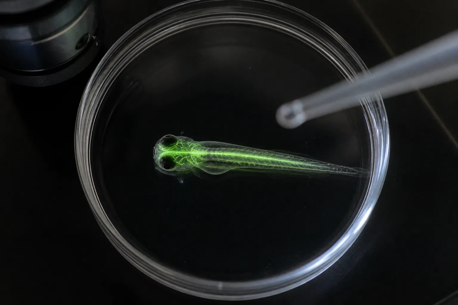
The nose was never the whole story. For decades, olfactory receptors were treated as a closed system: molecular sentinels stationed in the nasal epithelium, with each sensory neuron expressing one receptor type and each receptor tuned to a limited region of chemical space. That model remains useful for describing canonical smell, but zebrafish have exposed its blind spot.
In zebrafish, at least 36 olfactory receptor genes are active outside the classical olfactory tissues. Their expression has been detected in the head, trunk, pharynx and pancreas — places not usually described as sensory organs. The finding does not mean that every non-nasal receptor is detecting an odor in the ordinary sense. It means that the molecular machinery associated with olfaction is being reused in tissues where chemical information may serve other purposes.
That distinction is central to understanding non-olfactory olfactory receptors in zebrafish sensory systems. The important question is no longer simply where a receptor is expressed. It is what kind of signal the receptor encounters there, which G-proteins and effectors are available, and how the surrounding tissue interprets the resulting activity.
1. The GPCR landscape: four chemosensory families, four ways of reading chemistry
Olfactory receptors belong to the G-protein-coupled receptor, or GPCR, superfamily. They sit in the cell membrane and convert the presence of a chemical ligand into an intracellular signal. That basic architecture is shared across many receptor systems, but the sensory information extracted from it depends on the receptor family, the ligand environment and the cells that receive the signal.
In zebrafish, the main chemosensory GPCR repertoire is commonly divided into four families:
| Family | Full name | Typical sensory emphasis | Why it matters in zebrafish |
|---|---|---|---|
| OR | Odorant receptors | Detection of a broad range of odorant molecules | Provides the largest and most familiar part of the olfactory receptor repertoire |
| TAAR | Trace amine-associated receptors | Amines and volatile chemical cues often linked to biologically meaningful states | Adds sensitivity to a more specialized chemical space |
| ORA | V1R-related receptors | Pheromone-like cues and selected amino-acid signals | Includes a relatively small, distinct receptor group; zebrafish have seven ORA genes |
| OlfC | V2R-related receptors | Water-soluble odorants and social or environmental cues | Particularly important in an aquatic environment, where molecules are encountered in solution |
The four families are not interchangeable. Their ligand-binding properties and downstream signaling relationships give them different jobs within the broader chemosensory system. A zebrafish does not experience its environment through a single undifferentiated pool of “smell receptors.” It samples water through receptor populations that divide chemical space in different ways.
The aquatic setting is especially important. In air-breathing vertebrates, many familiar odorants reach the nasal epithelium as volatile molecules. A zebrafish encounters its chemical environment in water, where compounds dissolve, diffuse and form gradients through the surrounding medium. That makes water-soluble detection a primary design problem rather than a secondary adaptation. The expanded importance of the OlfC, or V2R-related, family fits this context.
This is also why receptor distribution cannot be interpreted without considering the medium around the animal. A receptor that appears unusual from a mammalian, nose-centred perspective may be entirely coherent in a fish. The chemical landscape is different, so the sensory architecture is different.
A receptor’s location does not define its identity. It defines the assumptions we are tempted to make about its function.
The GPCR landscape provides the first of five ways these receptors shape sensory input: it determines which kinds of chemical variation can be converted into neural or physiological signals in the first place. The receptor family is not just a label in a genome annotation. It is part of the system’s input filter.
2. Ectopic expression: when an olfactory receptor is not serving the nose
The most counterintuitive result from zebrafish expression studies is the presence of olfactory receptor transcripts in tissues with no obvious olfactory role. At least 36 olfactory receptor genes have been detected outside the nasal cavity and classical olfactory structures. The reported sites include the trunk, pharynx and pancreas.
“Ectopic” describes the location relative to the receptor’s canonical tissue. It does not automatically describe the receptor as abnormal, accidental or non-functional. In developmental biology, a gene can be active in several tissues while contributing to different processes in each one. The same receptor protein may participate in odor coding in an olfactory sensory neuron and in local chemical signaling elsewhere.
There are several non-exclusive explanations for this distribution.
Local chemical monitoring
Non-nasal tissues are chemically active environments. They contain metabolites, hormones, ions, nutrients and locally released signaling molecules. A receptor capable of detecting a small molecule in the nose could, in principle, provide a way for another tissue to monitor its own chemical surroundings.
The signal would not need to become a conscious sensation. A pancreatic cell, for example, could use a receptor-linked pathway to modify secretion or metabolism. A pharyngeal cell could respond to compounds passing through the digestive or respiratory interface. A cell in the trunk could use receptor activity as part of a local developmental or physiological program.
This is the key difference between sensory detection and sensory perception. A receptor can detect chemistry without producing an experience that an animal would recognize as smell.
Distributed physiological regulation
Ectopic olfactory receptors may also act as peripheral chemosensory nodes. Instead of transmitting information to the olfactory bulb, they may influence local tissue behaviour or connect chemical conditions to broader physiological states.
The possible outputs are not limited to neuronal firing. GPCR signaling can alter intracellular messengers, enzyme activity, ion channels, secretion and gene expression. The same receptor family therefore becomes capable of participating in regulation that is metabolic, endocrine or developmental rather than strictly neural.
This possibility is particularly relevant in organs whose main role is already defined in chemical terms. The pancreas is not a sensory organ in the conventional anatomical sense, but it constantly interprets the chemical state of the organism. Expression of olfactory receptor genes there suggests that chemosensory transduction mechanisms may be embedded within ordinary physiological regulation.
Developmental patterning
A third possibility is that some receptors are involved in tissue formation. During development, GPCR activity can influence cell movement, differentiation and communication between neighbouring cells. An olfactory receptor expressed in a developing tissue may therefore be contributing to morphogenesis rather than monitoring an external cue.
This interpretation needs to be handled carefully. Expression alone does not establish a developmental function. It does, however, make a purely nose-centred interpretation inadequate. If a receptor appears before a tissue is mature, or in a location where no plausible odorant access exists, its developmental context becomes part of the biological question.
Expression is not proof of function
The distinction between transcription and function matters throughout this field. Detecting an olfactory receptor transcript tells us that the gene is active, but not necessarily that the receptor reaches the cell surface, binds a ligand or produces a meaningful physiological response. Functional validation requires additional evidence: protein localization, appropriate signaling machinery, ligand response, perturbation experiments or a measurable change in tissue behaviour.
That caution does not weaken the ectopic-expression finding. It makes the finding more precise. Zebrafish reveal a wider map of receptor gene activity; the next task is to determine which locations support active signaling and which represent transient, low-level or developmental expression.
The second way these receptors shape sensory input is therefore spatial. They expand the number of tissues able to sample chemical conditions, but they also force researchers to separate anatomical presence from demonstrated sensory function.
3. Developmental timing: the sensory network begins before the nose is finished
Zebrafish development offers an unusually clear view of how a sensory system is assembled. Their larvae are transparent, development proceeds rapidly, and the major stages of nervous-system formation can be followed with high temporal resolution.
The olfactory placode forms as an ectodermal thickening at approximately 17 hours post-fertilization. By around 32 hours post-fertilization, the tissue has invaginated to form the nares. The interval is short, but it contains several distinct biological transitions: precursor cells are specified, the sensory epithelium takes shape, neurons begin to differentiate and connections with the olfactory bulb are established.
This timeline changes how ectopic expression should be interpreted. If olfactory receptor genes are active in the trunk, pharynx or other peripheral tissues while the olfactory organs are still developing, then chemical signaling is not waiting for a mature nose. Peripheral and canonical olfactory systems may be constructed in parallel.
That does not prove that the larva is already using every one of these receptors as a functioning sensory device. It does show that receptor expression must be placed on a developmental timeline. A measurement taken at one larval stage may capture a system in transition rather than a stable adult-like arrangement.
Several developmental anchors help keep the interpretation grounded:
- Around 17 hpf: the olfactory placode appears as a thickening of ectoderm.
- Around 32 hpf: the placode invaginates to form the nares.
- After the initial organ formation: olfactory sensory neurons mature, extend axons and contribute to the organization of glomerular patterns in the olfactory bulb.
- During ongoing larval development: receptor expression, neuronal identity and circuit connectivity continue to be refined.
The sequence matters because receptor choice and tissue function are not independent of maturation. A receptor transcript found in an immature cell may reflect an early developmental state, while the same transcript in a mature sensory neuron may support stable signal detection. Comparing the two without accounting for developmental stage can make a transient pattern look like a permanent design feature.
Development also provides a possible explanation for why receptor genes appear outside the nose. A receptor could be used first as part of a local developmental program and later become associated with chemical sensing. Alternatively, ectopic expression could identify tissues that are already chemically responsive before the main olfactory circuit has reached maturity.
In both cases, the zebrafish model makes a broader point about sensory systems: they are built as distributed processes. The sensory organ, receptor repertoire and central circuit do not all become functional at the same moment. Chemical information can be available to developing tissues before the mature architecture is in place.
That is the third way non-olfactory olfactory receptors shape sensory input: they introduce chemical sensitivity into developmental windows where a conventional map of the olfactory system would predict little or no sensory activity.
4. Cross-modal integration: the olfacto-retinal centrifugal pathway
Olfactory information does not remain confined to the olfactory system. In zebrafish, one of the clearest examples is the olfacto-retinal centrifugal, or ORC, pathway. It links the olfactory bulb with the retina and provides a route through which olfactory state can influence visual processing.
The pathway involves neurons associated with the terminalis system and projections from the olfactory bulb to dopaminergic interplexiform cells in the neural retina. Its direction is important. This is not simply a route carrying visual information toward an olfactory centre. It is a centrifugal modulatory connection: activity associated with olfactory input can alter how the retina handles visual signals.
The effect is mediated through dopamine. Retinal dopamine influences circuits that include bipolar and horizontal cells, both of which contribute to the way the retina organizes contrast, luminance and temporal information. The precise outcome depends on the state of the circuit, but the principle is clear: chemical context can change the settings of early visual processing.
For a fish moving through a chemical gradient, this is biologically sensible. Olfactory information can signal food, danger, social context or a change in the surrounding environment. Vision then needs to be interpreted against that background. The same visual scene may require a different response when the water carries a predator-associated chemical cue than when the chemical environment is neutral.
This is not the same as saying that smell creates a complete visual interpretation. The ORC pathway is better understood as a gain-control or state-setting mechanism. It changes the conditions under which visual signals are processed rather than replacing the visual input itself.
The distinction is useful because “cross-modal integration” is often described too broadly. Integration does not always occur at a high-level brain centre after each sensory system has completed its own analysis. In zebrafish, the interaction can begin through direct connections between sensory circuits, including a pathway that reaches the retina.
A simplified comparison makes the architectural difference clearer:
| Feature | Nose-centred model | Distributed zebrafish model |
|---|---|---|
| Primary site of olfactory receptor activity | Nasal olfactory epithelium | Nasal epithelium plus several non-nasal tissues |
| Main output | Olfactory sensory neurons and bulb circuits | Local tissue signaling, olfactory circuits and modulatory connections |
| Relationship to vision | Often treated as a later integrative process | Can include direct olfactory influence on retinal processing |
| Interpretation of receptor expression | Closely tied to odor detection | Requires tissue, developmental and signaling context |
| Meaning of chemical input | External odor identity | External cues plus internal or local chemical state |
The ORC pathway supplies the fourth way these receptors shape sensory input: chemical information can alter another sensory system before signals are combined into a unified behavioural decision. Sensory systems are not sealed channels. They tune one another continuously.
5. Regulatory mechanisms: receptor choice, G-protein feedback and chromatin state
The canonical olfactory system has a demanding organizational problem. A sensory neuron must select a receptor identity that can be read reliably by the rest of the circuit. In the classic one-receptor-per-neuron arrangement, the neuron expresses one dominant olfactory receptor while alternative receptor genes are silenced.
That arrangement is not produced by a single switch. It depends on a combination of stochastic gene activation, receptor feedback, G-protein signaling and epigenetic repression. Together, these mechanisms determine which receptor a neuron presents to the olfactory circuit and how stable that identity remains.
Initial selection is probabilistic
Immature olfactory sensory neurons may begin with a permissive chromatin state in which more than one receptor locus can be explored. Low-level or transient expression provides the cell with a route toward receptor selection rather than fixing the identity in advance.
The initial step is therefore not necessarily a clean, immediate choice. It is a period of competition among receptor genes. What matters is which receptor becomes functionally established and able to engage the feedback system.
Receptor activity reinforces identity
Once a receptor is expressed and coupled to its signaling machinery, its activity can feed back onto gene regulation. In the model described for zebrafish olfactory neurons, G-protein βγ subunits participate in a signal that suppresses alternative receptor transcription.
This gives the receptor two roles. It is a detector of chemical ligands, but it is also part of the mechanism that stabilizes the neuron’s identity. The receptor helps determine which receptor genes remain available and which are shut down.
The logic is economical. A neuron does not need a separate identity-maintenance system entirely disconnected from its sensory machinery. Activity in the chosen signaling pathway can become the signal that closes the selection process.
Histone methylation closes alternative loci
Feedback alone would not be sufficient if receptor genes could immediately reactivate. Stable receptor choice requires a more durable form of repression. Histone methylation contributes to that stability by placing repressive marks on alternative receptor loci.
Marks such as H3K9me3 and H3K27me3 are associated with compacted chromatin and reduced transcriptional accessibility. In this state, alternative receptor genes are not merely inactive for the moment; they are placed in a regulatory environment that makes renewed expression less likely.
The result is an epigenetic lock. As the neuron matures and becomes integrated into the olfactory bulb, its receptor identity remains sufficiently stable for the circuit to interpret its activity consistently.
The regulatory sequence can be summarized without treating it as a rigid linear machine:
1. Immature neurons maintain access to a range of receptor loci.
2. One receptor begins to achieve functional expression.
3. Receptor-linked G-protein signaling provides feedback on gene choice.
4. Alternative receptor loci acquire repressive chromatin states.
5. The selected identity is maintained as the neuron matures and connects to the olfactory bulb.
Why ectopic tissues may follow different rules
This regulatory architecture also clarifies why receptor expression outside the nose is so informative. A pancreatic cell or a pharyngeal cell is not an olfactory sensory neuron. It may not use the one-receptor-per-neuron rule, and it may not have the same feedback circuitry or chromatin constraints.
An ectopic cell could express several receptor genes at once. It could activate them temporarily during development. It could use only part of the canonical signaling cascade, redirecting the output toward secretion, metabolism or tissue growth. The presence of an olfactory receptor gene therefore does not imply that the entire olfactory-neuron program has been transplanted into another organ.
This is the fifth way these receptors shape sensory input: regulation determines whether chemical information is represented as a stable sensory identity, a local physiological response or a transient developmental signal. The receptor’s function depends not only on the ligand and the tissue, but also on the gene-regulatory environment that controls when and how the receptor can act.
A distributed sensory system, not a misplaced gene list
The significance of ectopic olfactory receptor expression is not that zebrafish possess a collection of biological curiosities outside the nose. Its significance is that the same molecular toolkit can be deployed at several levels of organization.
In an olfactory sensory neuron, a receptor contributes to combinatorial odor coding. In a developing tissue, related signaling may influence cell state or morphogenesis. In a peripheral organ, receptor activity may provide local information about the chemical environment. Through the olfacto-retinal centrifugal pathway, olfactory state can even modulate how visual information is processed.
These possibilities should not be collapsed into one universal function. The term “olfactory receptor” describes evolutionary and molecular family membership; it does not guarantee an identical role in every tissue. Function has to be established in context.
For experimental work, that means asking several questions at once:
- Is the receptor transcript present in a defined cell type or only in a mixed tissue sample?
- Does the receptor protein reach the plasma membrane?
- Are the relevant G-proteins and downstream effectors expressed in the same cells?
- Does exposure to a candidate ligand produce a reproducible response?
- Does changing receptor activity alter tissue behaviour, development or circuit function?
- Is the expression stable in mature tissue or restricted to a developmental stage?
The answers separate a compelling expression pattern from a demonstrated signaling pathway. They also prevent a common interpretive error: assuming that anatomical location determines biological identity.
The zebrafish model is valuable precisely because it makes this error difficult to sustain. Transparent larvae reveal the timing of organ formation and circuit assembly. Conserved vertebrate genetics make it possible to connect receptor families with broader principles of sensory biology. The animal’s aquatic environment also highlights receptor families and signaling strategies that can be less visible in nose-centred mammalian models.
The result is a less tidy but more accurate picture of chemosensation. Sensory receptors are not confined to the organs that first gave them their names. Their distribution reflects a broader biological strategy: reuse a responsive molecular system wherever chemical information can guide development, physiology or behaviour.
The nose remains central to olfaction. It is simply not the boundary of olfactory receptor biology.