Olfactory bulb circuits before and after sensory deprivation
A single day of unilateral naris occlusion is enough to remodel part of the olfactory bulb. The first measurable change is not a wholesale shutdown of odor processing.
It is cell-type specific: the axon initial segment, or AIS, becomes shorter in axon-bearing dopaminergic interneurons of the glomerular layer, and those cells become less intrinsically excitable.
That distinction sets the correct baseline for analyzing olfactory bulb circuits under sensory deprivation. Input loss does not produce one uniform response. The circuit redistributes gain. Some cells reduce their firing capacity. Primary sensory synapses strengthen their glutamatergic drive. Mitral cell axons change on both sides of the brain, including the hemisphere that still receives sensory input. Newly generated interneurons become smaller but more excitable.
The practical conclusion is direct: an “after deprivation” map cannot be read as a damaged version of the “before” map. It is a recalibrated system.
Rapid structural remodeling: the 24-hour threshold
The axon initial segment is the control point for spike initiation. Its position, length, and molecular composition shape how easily a neuron converts synaptic and intrinsic voltage changes into an action potential. If the AIS shortens, the cell’s firing behavior can shift even when its dendrites, synaptic partners, and gross morphology appear unchanged.
After 24 hours of unilateral naris occlusion, this remodeling occurs selectively in axon-bearing dopaminergic interneurons in the olfactory bulb’s glomerular layer. The same short deprivation window reduces their intrinsic excitability. The circuit therefore modifies both structure and output capacity.
The specificity matters more than the speed.
AIS-negative dopaminergic cells do not show the same structural change. Excitatory mitral and tufted cells also remain structurally unchanged at the AIS under this brief deprivation condition. That rules out a simple model in which every neuron exposed to reduced odor input shortens its spike-initiation zone.
A useful before-and-after map should therefore separate at least four variables:
- Cell identity: dopaminergic interneuron, mitral cell, tufted cell, or another interneuron class.
- Axon status: whether the cell possesses an axon-bearing AIS that can undergo the observed remodeling.
- AIS structure: length and position, measured within the same anatomical reference frame.
- Intrinsic excitability: current threshold, firing response, and action-potential output under controlled stimulation.
If these variables are collapsed into a single label such as “olfactory bulb neuron,” the main result disappears. The deprivation response is not a property of the bulb in the abstract. It is a property of a defined cell population inside a defined circuit layer.
Twenty-four hours is not a recovery interval. It is a circuit-remodeling interval.
Why the glomerular layer responds first
The glomerular layer receives the first organized synaptic input from olfactory sensory neurons. Axons from receptor neurons converge into glomeruli, where they contact mitral and tufted cells alongside local interneurons. Dopaminergic interneurons regulate this early stage of processing. Their output can alter presynaptic release, local inhibition, and the gain applied to incoming odor signals.
A deprivation-driven reduction in dopaminergic excitability can therefore change the operating range of the input layer without eliminating transmission. The circuit may be reducing one form of inhibition while adapting other nodes to maintain balance. The direction of the final sensory effect cannot be inferred from the AIS result alone.
This is where many “before versus after” diagrams fail. They show fewer sensory inputs and then draw weaker activity throughout the bulb. The data support a more mechanical sequence:
1. Sensory input falls on one side.
2. A defined dopaminergic population shortens its AIS and reduces intrinsic excitability.
3. Other synaptic and axonal compartments adjust over different time scales.
4. The resulting network changes sensitivity and selectivity rather than simply losing all activity.
The molecular cascade that links input deprivation to AIS shortening remains unresolved. That gap should stay visible in any mechanistic account. The structural result is established; the exact signal chain that produces it is not.
Synaptic scaling under input loss
Longer-term sensory deprivation produces a second class of adaptation at the synapse. Primary sensory neuron terminals in the olfactory bulb show an increased probability of glutamate release. Quantal currents mediated through AMPA and NMDA glutamate receptors also rise.
This is a classic circuit problem: the system receives less patterned input, yet the remaining sensory synapses become more effective. The circuit does not respond to reduced input by turning every gain control down. It adjusts transmission to preserve functional drive.
The distinction between input quantity and synaptic efficacy is essential. Naris occlusion reduces access to odorants on the blocked side. It does not mean that every primary sensory neuron stops releasing transmitter, nor that the olfactory bulb becomes electrically silent. The remaining signals can be amplified at the synaptic interface.
AMPA and NMDA receptors contribute on different operational scales. AMPA receptor currents provide fast depolarizing drive. NMDA receptor currents contribute voltage-dependent and temporally extended excitation. An increase in both receptor-mediated components changes not only the size of individual synaptic events but also the way coincident activity is integrated by postsynaptic cells.
The result is a circuit with less external sampling but stronger transmission per effective event. That combination can preserve responsiveness while degrading the precision of the representation.
How to read the synaptic change
A robust analysis should separate three measurements that are often mixed:
| Measurement | What it isolates | What deprivation changes |
|---|---|---|
| Presynaptic release probability | The likelihood that an active sensory terminal releases glutamate | Increases after longer-term sensory deprivation |
| AMPA-mediated quantal current | Fast postsynaptic response to individual glutamate events | Increases, indicating stronger fast excitatory drive |
| NMDA-mediated quantal current | Slower, voltage-dependent excitatory component | Increases, changing temporal integration and coincidence sensitivity |
These are not interchangeable readouts. A larger postsynaptic current does not by itself prove that more sensory neurons are active. A higher release probability does not prove that odor coding is more selective. Each result describes one segment of the transmission chain.
For circuit mapping, the practical sequence is to isolate the compartment first, then quantify the change:
1. Presynaptic terminal: measure release behavior.
2. Postsynaptic receptor response: separate AMPA and NMDA components.
3. Cell output: record firing threshold and spike patterns.
4. Network representation: test receptive-field width and odor discrimination.
This sequence prevents a common error: using a synaptic gain change as a direct proxy for improved perception. The downstream representation can move in the opposite direction.
Bilateral compensation: the non-deprived side is not a control
Unilateral sensory deprivation creates an asymmetric input condition. It does not create an isolated lesion. Mitral cell axons change on both sides of the olfactory system. Their morphology, myelin thickness, and action-potential spiking patterns are altered in the sensory-deprived hemisphere and in the non-deprived hemisphere.
That bilateral response changes the logic of experimental controls. The untreated side may retain normal odor access, but it is not necessarily a biologically neutral reference. The brain processes the imbalance between sides. A change in one hemisphere can alter network state, interhemispheric coordination, neuromodulatory tone, or the way bilateral sensory evidence is compared.
The finding also expands the definition of plasticity. Plasticity is not limited to the synapse that loses input. It can propagate into axonal structure and conduction properties. Myelin thickness affects how action potentials travel along the axon. Changes in axonal geometry and myelination can alter timing, reliability, and the coordination of output across connected regions.
The exact evolutionary purpose of this bilateral compensation remains unresolved. It is not safe to assign a single adaptive explanation. The measured fact is narrower and more useful: unilateral sensory loss produces system-wide changes that include the opposite, non-deprived pathway.
Mapping the circuit before and after deprivation
A useful olfactory bulb map should contain more than glomerular positions and cell counts. It should register the operating parameters that determine signal flow.
For each hemisphere, track:
- The location and organization of sensory input zones.
- The identity and excitability of local interneuron populations.
- AIS structure in cell types known to remodel.
- Primary sensory synapse release probability.
- AMPA- and NMDA-mediated postsynaptic responses.
- Mitral cell axon morphology and myelin thickness.
- Action-potential firing patterns.
- Receptive-field width and odorant discrimination performance.
The bilateral result also demands matched analysis. Compare deprived and non-deprived hemispheres, but do not assume that the latter defines the pre-deprivation state. If the experiment includes a separate unmanipulated group, that group provides the cleaner baseline.
This distinction is especially relevant when interpreting sensory receptor signaling changes. Alterations in receptor neuron input, synaptic release, bulb interneuron excitability, and mitral cell output occupy different levels of the pathway. They should be mapped as linked stages, not merged into one broad claim about “olfactory sensitivity.”
Homeostatic balancing: smaller interneurons, higher excitability
Sensory deprivation also affects newly generated interneurons in the olfactory bulb. Their size decreases, but their intrinsic excitability increases. The two observations are not contradictory. They represent compensation across separate control variables.
A smaller cell can have different membrane properties, input resistance, dendritic integration, and electrotonic structure. Increased intrinsic excitability can offset some of the functional consequences of reduced cellular size. The system does not preserve morphology for its own sake. It preserves an operating range.
This is the core of homeostatic plasticity: the circuit adjusts several parameters in opposite directions to stabilize output. One compartment loses scale; another gains responsiveness.
That compensation should not be described as a complete restoration of normal function. The evidence supports a balancing response, not a return to the original state. A cell that is smaller and more excitable may not integrate inputs in the same way as a normally sized cell. It may reach threshold more readily but process spatial or temporal patterns differently.
The correct question is therefore not, “Did the neuron recover?” It is, “Which function did the circuit stabilize, and which function did it trade away?”
Structural and functional variables can move in opposite directions
When analyzing deprived tissue, classify each result as structural, synaptic, intrinsic, or network-level:
- Structural: AIS length, cell size, axon morphology, and myelin thickness.
- Synaptic: release probability and receptor-mediated quantal currents.
- Intrinsic: threshold, input-output relationship, and firing response to current injection.
- Network-level: receptive-field width, sensitivity, and discrimination.
Then test whether the variables move together. In this system, they do not:
- Dopaminergic AIS length decreases.
- Dopaminergic intrinsic excitability decreases.
- Newly generated interneuron size decreases.
- Newly generated interneuron intrinsic excitability increases.
- Primary sensory synaptic release increases.
- AMPA and NMDA receptor-mediated currents increase.
- Mitral cell axonal properties change bilaterally.
This pattern is not noise. It is distributed control. The olfactory bulb reallocates gain between cellular compartments.
A single summary score would hide that logic. A circuit that appears “more excitable” at the synaptic level may contain a specific interneuron population that has become less excitable. A circuit with stronger sensory synapses may still produce broader, less selective mitral cell responses.
Increased sensitivity, weaker discrimination
The network-level consequence of early sensory deprivation is a sensitivity–selectivity trade-off. In rodent olfactory bulbs, early deprivation widens mitral cell receptive fields while increasing overall odorant sensitivity. At the same time, fine odorant discrimination becomes worse than in normally stimulated bulbs.
This is the key functional result. Reduced sensory exposure does not simply make the system less responsive. It can make the bulb respond to more odorants while reducing the separation between odor representations.
A wider receptive field means that a given mitral cell responds across a broader set of odor conditions. Increased sensitivity means that weaker or less complete inputs can drive the system. These changes may preserve detection. They also reduce the sharpness of the code if neighboring odor representations become less distinct.
The architecture has shifted from precise filtering toward broader access.
The deprived bulb does not necessarily hear less. It distinguishes less cleanly.
This distinction matters for interpreting sensory deprivation neural plasticity. If an experiment measures only response amplitude, it may conclude that the circuit has compensated successfully. If it measures discrimination, the result may look impaired. Both findings can be true because they describe different outputs.
A practical performance matrix
| Circuit property | Before deprivation | After deprivation |
|---|---|---|
| Sensory input | Normal access to odorant-driven receptor activity | Reduced input on the occluded side |
| Primary synaptic transmission | Baseline release and receptor-mediated currents | Higher release probability and stronger AMPA/NMDA-mediated currents |
| Dopaminergic interneuron AIS | Baseline structure in axon-bearing cells | Shortened after 24 hours of unilateral occlusion |
| Dopaminergic intrinsic excitability | Baseline firing capacity | Reduced in the affected axon-bearing population |
| Newly generated interneurons | Larger morphology with baseline excitability | Smaller morphology with increased intrinsic excitability |
| Mitral cell receptive fields | Narrower odorant response range | Broader response range |
| Overall odorant sensitivity | Baseline detection profile | Increased sensitivity |
| Fine odor discrimination | More selective separation | Reduced discrimination performance |
| Axonal adaptation | Baseline morphology and myelin | Bilateral changes in morphology, myelin, and spiking |
The table is not a substitute for pathway-specific measurements. It is a control against category errors. “Sensitivity,” “selectivity,” and “excitability” should never be treated as synonyms.
What this means for sensory-system models
The olfactory bulb is often used as a compact model for sensory processing because receptor neurons converge into organized glomerular structures and then pass through defined interneuron and projection-neuron layers. Sensory deprivation exposes the limits of a static wiring diagram.
A static map can show where cells connect. It cannot show how those connections change their operating parameters after input loss. To model the deprived circuit, the map must include at least four forms of adaptation:
1. Spike-initiation remodeling. AIS structure changes in a restricted dopaminergic population within 24 hours.
2. Synaptic scaling. Primary sensory synapses increase release probability and strengthen AMPA/NMDA-mediated currents.
3. Axonal recalibration. Mitral cell morphology, myelin, and spiking change bilaterally.
4. Homeostatic compensation. Newly generated interneurons reduce in size while increasing intrinsic excitability.
These changes occur at different time scales and may pull the circuit in different directions. The early AIS response is fast and cell-specific. Synaptic and axonal adaptations extend the response across the network. Receptive-field and discrimination changes describe the final computational output.
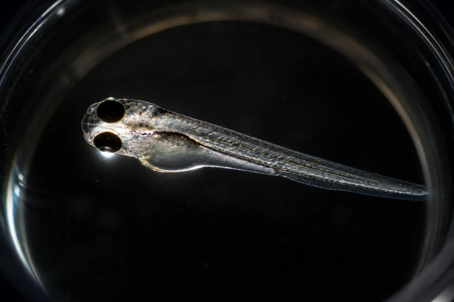
The sequence also clarifies how to approach comparative work, including studies of olfactory deprivation in zebrafish. The underlying logic may be useful across species, but the measurements cannot be transferred without validation. Rodent olfactory bulb findings should not be presented as direct zebrafish results. Species differ in anatomy, receptor organization, bulb circuitry, developmental timing, and the balance between olfactory and other chemosensory pathways.
For zebrafish experiments, the right move is to test the corresponding variables rather than import the conclusions:
- Does sensory blockade alter AIS structure in the same cell class?
- Does primary sensory release increase?
- Do AMPA and NMDA components scale together?
- Are axonal and myelin changes bilateral?
- Do receptive fields widen?
- Does detection improve while discrimination declines?
That is a comparative framework, not a forced equivalence.
A troubleshooting protocol for circuit analysis
When an olfactory bulb deprivation result looks inconsistent, isolate the failure point before explaining the biology. Use this sequence:
1. Confirm the deprivation geometry. Unilateral naris occlusion is not equivalent to complete sensory silence or bilateral deprivation. Record the side, duration, and developmental stage.
2. Separate cell classes. Do not combine axon-bearing and AIS-negative dopaminergic cells. Do not extend the 24-hour AIS result to mitral or tufted neurons.
3. Split morphology from excitability. A shorter AIS, a smaller cell body, and a lower firing threshold are different measurements. Quantify each independently.
4. Split pre- and postsynaptic effects. Increased glutamate release and larger AMPA/NMDA currents identify different adaptation sites.
5. Analyze both hemispheres. Treat the non-deprived side as an active biological condition, not an automatic baseline.
6. Measure sensitivity and discrimination separately. A stronger response to odorants can coexist with poorer fine discrimination.
7. Keep the time axis explicit. The 24-hour AIS response, longer-term synaptic scaling, bilateral axonal changes, and early receptive-field effects should not be merged into one undated condition.
8. Mark unresolved mechanisms. The molecular cascade producing dopaminergic AIS shortening is still unknown. State the structural result without inventing the pathway.
This protocol keeps the analysis aligned with what the circuit actually does. It also prevents the most common overstatement: treating sensory deprivation as a simple loss of function.
The circuit after deprivation is recalibrated, not erased
The olfactory bulb responds to missing input by redistributing control across its wiring. Dopaminergic interneurons alter the geometry of spike initiation within a day. Primary sensory synapses strengthen their glutamatergic transmission. Mitral cell axons remodel on both sides. Newly generated interneurons become smaller but more excitable. At the behavioral-output level, sensitivity rises while fine discrimination weakens.
That is a coherent system response. It preserves access to sensory signals, but it changes the cost of that access. The bulb becomes less selective in exchange for broader responsiveness.
Any credible before-and-after model should therefore do three things: identify the cell type, locate the altered compartment, and measure the resulting computation. If those parameters are missing, the map is incomplete. If they are present, sensory deprivation becomes readable as a sequence of structural and functional recalibrations rather than a generic story of neural decline.