Sensory integration: 3 ways to map taste and smell
When you bite into a ripe strawberry, your tongue does not taste "strawberry." It registers a pattern of sugars, acids, and faint bitter notes.
The fruit's signature — that bright, floral, sun-warm fragrance — arrives through the back of the throat, where volatile compounds travel retronasally to reach the olfactory epithelium. What you experience as flavor is not a property of the food. It is a multisensory construct assembled by the brain, and mapping how that construct forms is one of the most intricate problems in modern sensory neuroscience.
The challenge is not that the brain lacks regions for this work. It is that the relevant regions — the gustatory cortex, the piriform cortex, the orbitofrontal cortex, the anterior insula — do not behave as independent modules. They integrate signals continuously, weighting inputs dynamically, and the weights shift with hunger, context, and learning. To chart this convergence, researchers have developed three complementary families of sensory integration mapping methods: functional neuroimaging in humans, single-cell electrophysiology in behaving animals, and spatial transcriptomics combined with single-cell RNA sequencing. Each illuminates a different layer of the same circuit.
The architecture of flavor: where two streams converge
The taste pathway begins at the periphery. Receptor cells on the tongue and palate transduce five canonical qualities — sweet, salty, sour, bitter, umami — and relay signals through cranial nerves VII, IX, and X to the nucleus of the solitary tract (NST) in the brainstem. From there, the signal ascends to the thalamus and onward to the primary gustatory cortex, located in the insula.
Olfactory information travels a parallel but distinct route. Odorants bind to G-protein-coupled receptors in the olfactory epithelium, activate a canonical cAMP signaling cascade, and trigger neural impulses that project through the cribriform plate to the olfactory bulb. From the bulb, mitral and tufted cells relay the signal to the piriform cortex — the largest target of olfactory input — without an obligatory thalamic relay. This direct pathway allows olfactory signals to reach association cortex faster than taste signals, which is one reason smell so powerfully shapes the first impression of a food.
The two streams converge in higher-order association areas, particularly the orbitofrontal cortex (OFC), the anterior cingulate cortex (ACC), and the dorsal insula. In these regions, gustatory and retronasal olfactory signals combine to encode subjective pleasantness and food reward value. The OFC, in particular, acts as a secondary association area where associative learning binds specific taste-odor pairings to their hedonic consequences.
Flavor lives in the convergence, not in the tongue or the nose — it is a multisensory construct the brain builds from two parallel sensory streams.
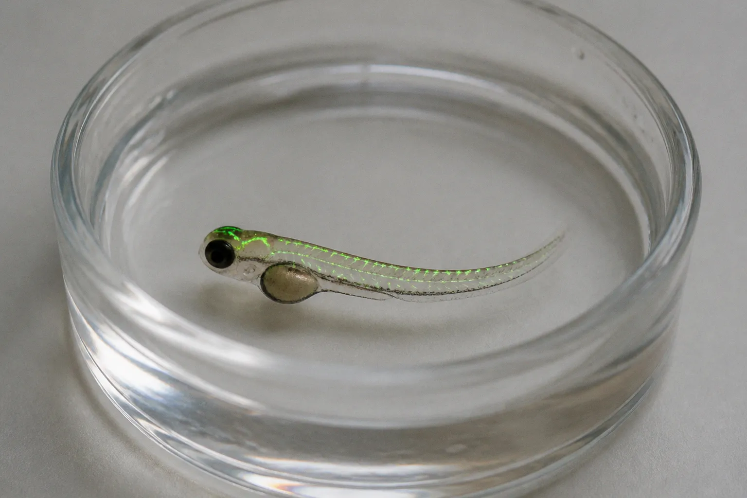
This organizational logic is broadly conserved across mammals, and similar chemosensory principles appear in zebrafish, where transparent larvae offer whole-brain imaging at single-cell resolution and a genetic toolkit that no mammalian model can match.
Method 1: Functional neuroimaging — mapping convergence in humans
The most direct window into human flavor processing is functional magnetic resonance imaging (fMRI). When a congruent taste-odor pair — sweet taste paired with a sweet-associated aroma, for instance — is delivered retronasally, brain regions involved in both taste and smell show superadditive responses: the activation is greater than the sum of responses to each component presented alone. This pattern has been consistently documented in the anterior cingulate cortex, the dorsal insula, and the orbitofrontal cortex.
What fMRI reveals is the spatial map of integration — where in the human brain taste and smell signals converge and amplify. The technique also captures how the OFC encodes subjective pleasantness. The same appetitive odor delivered to a recently sated subject produces weaker OFC activation than to a hungry one, showing that the neural integration is not purely sensory but deeply modulated by internal state, expectation, and learned association.
The method's limitation is resolution. fMRI measures hemodynamic responses, not neuronal spikes, and the signal is averaged across millions of neurons and seconds. It can tell you that a particular region is more active when a congruent taste-odor pair is perceived as pleasant, but it cannot identify which neurons are doing the work or how individual cells weight taste versus olfactory input moment by moment. It is also constrained to correlational observations: brain activity and behavior are recorded together, but causation must be inferred from indirect manipulations or follow-up studies in animal models.
Method 2: Single-cell electrophysiology — decoding cross-modal neurons
To capture the neural code at single-cell resolution, researchers turn to in vivo electrophysiology in behaving rodents, and increasingly in zebrafish. Microelectrodes or multi-electrode arrays record the action potentials of individual neurons while the animal samples taste, odor, or combined taste-odor stimuli delivered through intraoral cannulas.
The central finding from this approach is that chemoselective neurons in the gustatory cortex and posterior piriform cortex exhibit nonlinear cross-modal responses to taste-odor mixtures. A single neuron may respond to sweet taste, to a specific odor, and to the combination — but the combination response is not predictable from the sum of the unimodal responses. Some neurons show enhancement, others suppression, and some fire only when the two inputs are presented together. This is the cellular signature of multisensory integration, and it reveals that the brain's flavor code is not a simple addition but an emergent property of convergent inputs.
A neuron that responds to sweet and to vanilla individually may respond to the combination with a third, entirely different firing pattern — the brain's flavor code is emergent, not additive.
Electrophysiology offers millisecond temporal resolution and single-cell specificity, but it is limited in spatial coverage. Recording from hundreds of neurons across many brain regions simultaneously remains technically demanding, and the technique is invasive — typically restricted to animal models. In zebrafish, the method takes a different form: calcium imaging of genetically encoded indicators allows researchers to record from thousands of neurons simultaneously while the animal behaves, bridging the gap between cellular precision and brain-wide coverage. This makes zebrafish a particularly attractive model for multisensory processing research, where the goal is to relate the activity of defined cell classes to the structure of the surrounding circuit.
Method 3: Spatial transcriptomics — charting the molecular landscape
The newest addition to the sensory mapping toolkit is molecular. Spatial transcriptomics combined with single-cell RNA sequencing allows researchers to profile gene expression in individual cells while preserving their spatial location within the tissue. In a landmark study, this combination enabled the mapping of over 5.5 million neurons across more than 300 mice, revealing the spatial organization of olfactory receptor expression from the nose to the olfactory bulb.
What transcriptomics reveals is not activity in real time but molecular identity and circuit architecture. Researchers can determine which receptor types are expressed in which neurons, how those neurons are organized spatially, and how the molecular map relates to functional connectivity. The technique has confirmed that more than 1,000 types of olfactory receptors are organized in spatial bands across the olfactory bulb, a finding that bridges molecular biology with systems-level neuroscience and provides the substrate upon which sensory experience is built.
The limitation is that transcriptomics is largely a snapshot. It excels at structure and cell-type classification but does not directly capture dynamic processing. To link molecular identity to functional role, researchers must integrate transcriptomic data with electrophysiology or imaging — a hybrid strategy that is rapidly becoming the standard for chemosensory mapping techniques.
At a glance: strengths and limitations
| Parameter | fMRI | Single-cell electrophysiology | Spatial transcriptomics + scRNA-seq |
|---|---|---|---|
| Organism | Humans | Rodents, zebrafish | Mice, other models |
| Resolution | ~3 mm spatial, ~1 s temporal | Single neuron, ~1 ms temporal | Single cell, molecular |
| What it measures | Hemodynamic (BOLD) activity | Action potentials | Gene expression, cell identity |
| Strength | Whole-brain coverage in humans | Real-time single-cell precision | Comprehensive molecular atlas |
| Limitation | Indirect, low spatial precision | Invasive, limited coverage | Static snapshot, no dynamics |
| Key insight | Superadditive responses in ACC, insula, OFC | Nonlinear cross-modal coding in single neurons | Spatial organization of 1,000+ receptor types |
The three methods answer different questions. fMRI asks: where in the human brain do taste and smell converge? Electrophysiology asks: how do individual neurons encode the combination? Transcriptomics asks: what is the molecular architecture that makes this convergence possible? A complete picture of taste and smell integration requires all three, and the most informative recent studies are those that combine them.
What remains unmapped
Despite these advances, significant gaps remain. The full cellular-level mechanism by which human cortical networks dynamically re-weight taste versus smell during real-time consumption is still unmapped. Whether the primary gustatory cortex is organized by taste quality in a strict spatial map — gustotopy — or relies on ensemble temporal coding remains an active debate. And the trigeminal system — the chemosensory pathway responsible for the burn of chili, the cool of menthol, the tingle of carbonation — is often overlooked in flavor studies despite its substantial contribution to food perception and its direct relevance to receptor signaling beyond the classic five tastes.
Zebrafish offer a particularly powerful bridge across these scales. Their larvae are transparent, genetically tractable, and possess a conserved chemosensory system. Researchers can image entire brains at single-cell resolution while the animal responds to taste and odor stimuli, effectively combining the strengths of all three methods in one organism. As the genetic and optical tools for zebrafish continue to mature, this model is likely to play an expanding role in chemosensory mapping, particularly for questions about how sensory maps are established during development and how they are shaped by experience.
The principle behind the map
Mapping sensory integration is not a matter of choosing one technique. It is a matter of recognizing that flavor perception is a layered phenomenon — molecular, cellular, and systems-level — and that each method reveals a slice of the same circuit. The practical takeaway for researchers, students, and curious readers: match the method to the question, and integrate across methods whenever the data allow. The brain does not respect disciplinary boundaries, and the most accurate map of taste-smell integration will come from methods that talk to each other.