Mapping the Complete Architecture of Spinal-Projecting Neurons in Larval Zebrafish
A new eLife study maps spinal-projecting neurons across the entire larval zebrafish brain, giving researchers their first whole-brain view of the descending motor architecture.
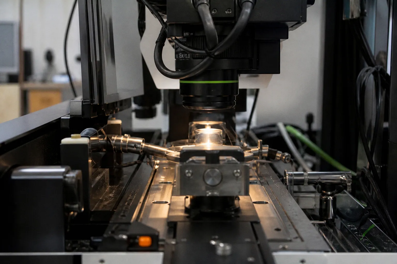
The work, published August 24, 2026, targets a long-standing bottleneck: how command signals travel from brain to spinal cord during behavior. For anyone running zebrafish circuit labs, the dataset reframes what can be measured at the systems level.
What the map delivers
The project isolates every neuron projecting from brain to spinal cord and registers each cell within a shared anatomical framework. The analytical step is straightforward: take sparse, hard-to-target projection neurons, assign them to brain regions, and quantify the spatial distribution. That conversion turns a qualitative tracing problem into a reproducible atlas entry. The payoff is concrete. Motor command pathways can now be compared animal-to-animal without re-running sparse injections from scratch.
Where the broader signal sits
The mapping arrives alongside two computational pieces published the same week. A Neural Computation paper from August 22 proposes a framework for quantifying information stored directly in synaptic connections rather than in firing activity — a useful complement when interpreting stable connectivity maps like the zebrafish atlas. Two days later, PLOS Computational Biology reported that manifold-constrained plasticity keeps recurrent networks stable during learning, reinforcing the case that wiring structure, not spike patterns alone, carries the durable signal. Together, the three outputs shift emphasis from activity to architecture — a methodological tilt worth noting in any grant draft or methods section.
What to check before citing
Before pulling the atlas into your own work, verify three parameters. First, confirm the developmental stage — larval mapping does not transfer cleanly to juvenile or adult circuits. Second, isolate the tracing method used; retrograde labeling versus genetic driver lines produces different cell-type coverage. Third, calibrate against your own registration pipeline; coordinate frames vary between atlases, and downstream comparisons fail silently if you skip the alignment audit. Keep the depression-neurogenesis ScienceDaily item on the radar only as adjacent context — it sits outside the circuitry focus and should not be folded into the same discussion.