New High-Speed Microscopy Captures Real-Time Electrical Activity Across the Zebrafish Brain
According to MIT News, a team at MIT has built a microscope that captures voltage fluctuations across an entire zebrafish brain at sub-millisecond timescales, with the work published in Nature Methods.
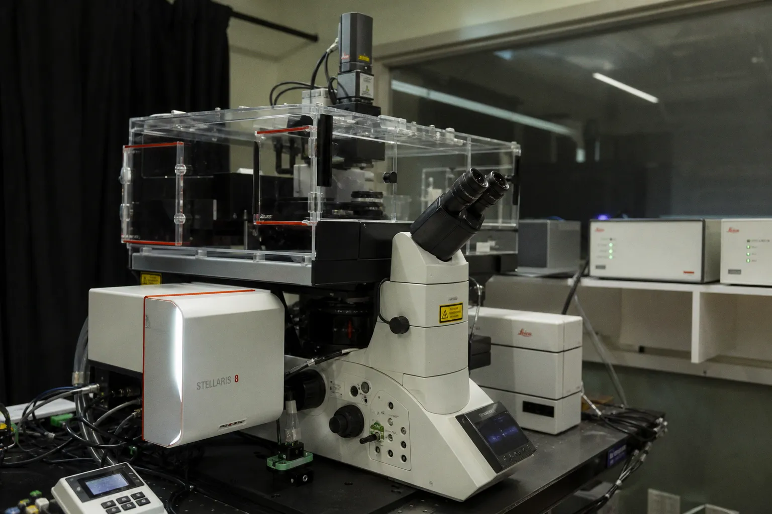
MIT engineers have unlocked a direct window into brain-wide electrical activity. According to MIT News, a team at MIT has built a microscope that captures voltage fluctuations across an entire zebrafish brain at sub-millisecond timescales, with the work published in Nature Methods.
The bottleneck: calcium imaging runs too slow
Standard circuit mapping relies on calcium sensors. Calcium floods into neurons after they fire, so it serves as a proxy for activity. The problem is timing. Calcium signals linger for seconds. Spike trains unfold in milliseconds. You lose the rhythm.
Jie Zhang, one of the lead authors alongside Zeguan Wang PhD '24, puts the gap plainly: neurons compute with voltage, not calcium. Direct voltage observation is the only way to see the actual signal, not a delayed echo of it. The old proxy treated each spike as a blur; the new method treats it as a discrete event.
The method: voltage indicators plus a fast swept microscope
The MIT group paired genetically encoded voltage indicators—fluorescent proteins that brighten when a neuron fires—with a microscope adapted for high-volumetric-rate imaging. The setup captures electrical activity across the whole brain on millisecond timescales.
In their demonstration, UV light triggered neurons throughout the zebrafish brain. The new scope recorded the resulting activity patterns across the entire volume. Now you can map which populations co-activate when a stimulus hits, not just where calcium pools minutes later.
Ed Boyden, the senior author, frames the goal in circuit terms: if you want to understand how neurons work as an emergent whole, you need to see them function as an emergent whole. Whole-brain, real-time capture is the prerequisite for that analysis.
What to track for your own imaging pipeline
Four parameters to calibrate before adopting the approach:
- Temporal resolution. Confirm the voltage indicator's response kinetics match your target spike train frequency. Sub-millisecond capture is only useful if the fluorophore can keep pace.
- Volumetric coverage. The technique prioritizes whole-brain sampling over single-cell resolution. You trade somatic detail for circuit-level synchrony. Quantify that trade-off for your experiment.
- Indicator expression. Genetically encoded voltage indicators require proper delivery—viral vectors, transgenic lines, or electroporation. Verify expression uniformity across the regions you intend to compare before drawing network conclusions.
- Behavioral compatibility. The scope is built for living animals. Validate that mounting, illumination, and indicator load do not distort the behavior you are trying to correlate with neural activity.
The headline win: you can now read electrical activity directly, across the whole brain, in behaving zebrafish. For labs mapping circuit development or linking neural activity to behavior, this shifts the baseline from inference to measurement.