Mapping Whole-Brain Responses to Nutrient Intake in Larval Zebrafish
If you've ever tried to pin down whether that one brain region you love is actually doing what you think it's doing during a feeding assay — and your honest answer is "eh, maybe, the data's too…
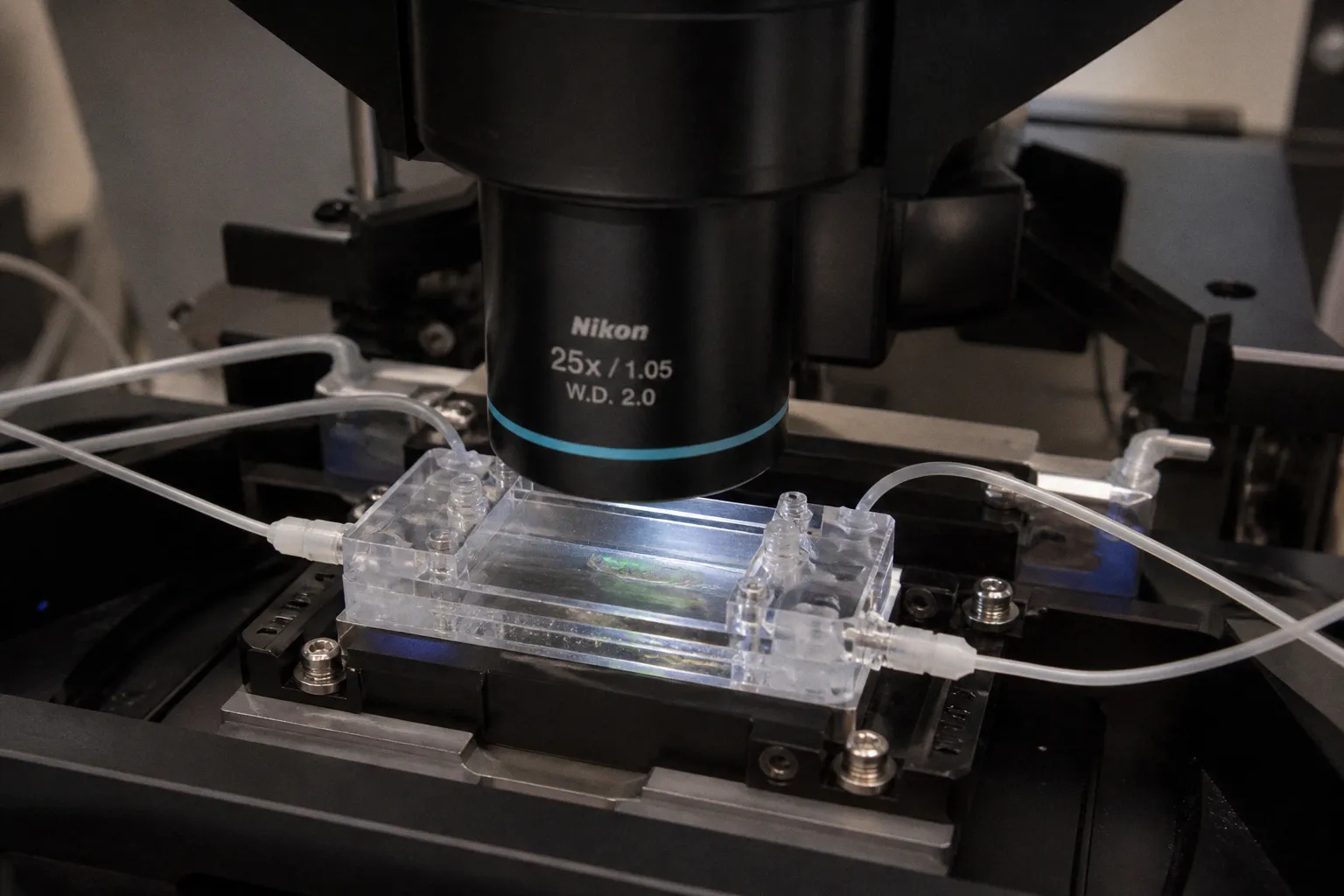
If you've ever tried to pin down whether that one brain region you love is actually doing what you think it's doing during a feeding assay — and your honest answer is "eh, maybe, the data's too noisy" — go read the new Nature Communications paper that just landed. Researchers at Janelia Research Campus (Howard Hughes Medical Institute), led by Misha B. Ahrens and James E. Fitzgerald, have stitched together an all-optical rig that lets you image whole-brain activity in behaving larval zebrafish while delivering nutrients straight to the gut or into the bloodstream, on top of standard visuo-motor stimulation.
What they actually built
The team — with Weiyu Chen, Ben James, and Virginie M. S. Ruetten as co-first authors — wired a light-sheet imaging path into a UV uncaging laser with a custom-built attenuator so they could release amino acids or D-glucose (but not the metabolically inert L-glucose) into either the gut or the vasculature, all while running the same larvae through visuo-motor stimulation. Everything stays all-optical: no pulling the fish out of the rig mid-trial, no need to switch modalities between imaging and physiology. You prep your larvae, you mount them, you image the whole brain at cellular resolution, and you perturb body chemistry in the same session. That's the closed-loop setup most of us have been sketching on whiteboards for the better part of a decade.
What the brain actually does
Here's the headline: nutrient signals light up neurons across the entire brain — well beyond any single feeding area — and the activity unfolds on multiple timescales spanning peripheral and central regions. Crucially, the evoked response depended on where the nutrient was delivered, which gives you a built-in anatomical control. Many gut-responsive neurons also fired during swimming and during visual stimulation, but the integration was neatly hierarchical. Brainstem regions mainly fused gut and motor signals; midbrain regions pulled together gut, visual, and motor inputs. In other words, the body-brain axis most labs have only inferred from sparse recordings is now on the table as a brain-wide, cellular-resolution map in an awake, behaving vertebrate.
Why this matters for your bench
If your work touches interoception, ingestive behavior, or sensorimotor integration, this is the platform you've been waiting for. You can borrow the optical uncaging tricks to ask whether your gene of interest changes gut-to-brain signaling, or use the brainstem-versus-midbrain split to figure out which level of the hierarchy your manipulation is actually hitting. One non-negotiable for your own prep: don't skip the D-glucose versus L-glucose comparison. Bake it in from day one, because it's the cleanest argument you'll ever make that you're engaging genuine nutrient-sensing pathways and not just stressing cells with a sugar puff. Go read the paper, then start sketching how you'd bring it to your own fish.